Microbes and me
Biology in Middle Schools home | |Elementary School sister project
Biology In Middle Schools is a Saint Michael's College student project. Link under 'toolbox' for a printer-friendly version. Click on handouts to print full resolution versions. Please see Wikieducator's disclaimer, our safety statement, and the Creative Commons licensing in English and in legalese.
Primary biological content area covered
- Prokaryotic vs. eukaryotic cells
- Diversity and ubiquity of prokaryotic life
- Interactions of people and microbes
- normal microflora
- microbes in food
- health consequences of disruptions of normal microflora
- basic mechanism(s) of infection
Materials
Rubber gloves a good idea for everyone
- Required for teacher
- Required for each student group
- Required for each student
- Prepared sterile petri dish
- sterile swabs
- Sterile water
- Tape
Description of activity
The students will start by learning basic facts about prok. and euk cell structure (ex. cell wall,basic organelles, placement of genetic material, basic metabolism). Next they will learn some prokaryotic facts, such as how some are healthy to have in and on your body, but can also cause disease in other circumstances. Also they will learn basic sterile technique which will be used during the activity.
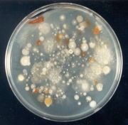
For an activity the students will be allowed to test what they have been taught. They will swab an object in the room, and let the bacteria grow on an agar plate (figure 1) over a period of a week. This activity is designed to demonstrate that microbes can be found in common places (just about any and everywhere) around the classroom. To avoid especially harmful bacteria, students should not be allowed to swab any orifices, bodily fluid or bathroom. Also the students will be able to see how the microbes create colonies over a period of time.
Briefly describe the activity, but provide enough detail so that the activity can easily be assessed by other teachers without your intimate knowledge of the topic.
Lesson plan
- Part 1: Background discussion
- Prokaryotes vs. Eukaryotes
- defined by presence or absence of membrane-bound nucleus – prokaryotes with, eukaryotes without
- prokaryotes still have DNA, arranged in a single, circular chromosome - instead of nucleus, area in bacterial cell where DNA is located called nucleiod
- prokaryotes also lack other internal membranes, things like mitochondria, chloroplasts (in plants), endoplasmic reticulum, Golgi apparatus, etc. – can talk about these organelles according to students knowledge from other coursework.
- forms of life in each group: bacteria (Kingdom Monera) comprise prokaryotes, eukaryotes all others: plants, animals, fungi, protists
- all prokaryotes are single-celled organisms-there are also single-celled eukaryotes (yeast are a familiar example), but any multi-celled organism is automatically eukaryotic)
- cell walls are found in both groups, but only some eukaryotes have cell walls (e.g. plants, fungi). Different cell walls composed of different substances:
- Plants: cellulose (what we know as dietary fiber)
- Fungi: chitin
- Bacteria: peptidoglycan
- Characteristics of Prokaryotes (Bacteria)
- remember that each cell constitutes a separate organism
- have the most diverse metabolisms of any kingdom
- different bacteria can use almost any energy source, tolerate any temperature range, and thus can live in the most diverse habitats – from your skin to the ocean floor to hot springs to dirt, you name it.
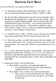
- live in huge numbers – a fun fact for the students: there are more bacteria in a spoonful of soil than the number of people who have ever lived (6.7 billion people on Earth right now)
- bacterial cell walls are one of two major types, called Gram-positive and Gram- negative based on what color they stain after a specific staining procedure (called a Gram stain)
- Bacteria and Human Health
- bacteria can be helpful or harmful to human health
- body always has normal microbial populations, both inside and out
- skin is always populated by normal bacteria – provides competition for pathogenic (disease-causing) bacteria. Staphylococcus species are common on skin
- intestines have bacteria that aid in digestion (E. coli, for example) Useful Web Site
- bacteria become a problem when they significantly increase in number and/or when they colonize normally sterile areas – blood is the best example of this – bacteria that enter the blood can travel all over the body and infect every major organ.
- because bacteria are everywhere, and because they grow very easily and can cause serious illness, it’s important to be careful about infection and take the proper precautions to prevent the spread of bacterial infections – good hygiene, clean bathrooms, etc – also why sterile technique is important in laboratory exercises. People can be exposed to bacteria from virtually any source – food, household surfaces, healthcare settings (could mention MRSA) unclean wounds (soil), etc.
- Prokaryotes vs. Eukaryotes
- Part 2: Lab
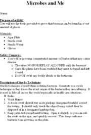
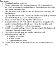
- The students will receive a handout with the protocol on it (figures 2-3). The information is repeated here, with slight modifications/additions for teacher reference.
- Sterile technique:
- Sterile technique is used when isolating bacteria. Scientists use sterile technique so they know the exact origin of the bacteria they are culturing. It is used in labs all across the world especially in health care situations.
- Rules
- Wash Hands! Make sure students
- A sterile swab should stay in its package (unopened) until it is used for testing. It should only touch the object being tested, then be disposed of in a designated garbage bag.
- Keep petri dishes closed until testing. Open it slightly so you can rub the swab on the agar, and quickly re-cover. This keeps airborne bacteria from growing on the plate.
- Procedure:
- Wash hands and put on gloves
- Discuss with partner what area in class room will be interesting to test. Note: Areas that are touched often ex. Keyboards and doorknobs will contain a lot of bacteria.
- Take out sterile swab (make sure the end doesn’t touch anything but the test object)
- Dip swab into sterile water. Water will help the swab pick up bacteria from the test object, and pass it onto the agar plate.
- Rub moist swab all over the object for a few seconds.
- To transfer bacteria, rub swab all over the agar (use enough force to transfer bacteria, but do not break agar). Keep the top of the plate over agar to avoid airborne bacteria entering agar.
- Quickly close plate and tape closed. It is important to label plate with your names, date and location tested.
- Place plate in a warm area, and wait for bacterial growth. Without a dedicated incubator, this could take from a day to a week depending on conditions
- Make observations each day
- Number of colonies
- Colony Colors
- Approximate size (mm)
- Discuss which areas had the most bacteria and possible sources of error. Make sure the students understand that each colony represents what was originally a single bacterial cell that divided into millions of cells, visible on the agar plate as a colony; thus, counting colonies (if there are distinct colonies and not a bacterial “lawn”) is theoretically equivalent to counting the number of cells plated.
Potential pitfalls
- Health concerns - potential exposure to potential pathogens
- keep plates closed after initial streaking
- rubber gloves
- Lack of incubator
- will grow at room temp, just slower - not a single class period activity
- potential to be an overwhelming topic - emphasis on simplicity and basic concepts is key
Math connections
- Include brief discussion of culture growth Useful Web Site
- Describe exponential Growth (and growth curves) compared to linear growth
- simple calculations of culture densities to incorporate multiplication, densities, etc. as desired by teacher
Connections to educational standards
- S7-8:30 (DOK 2)
- Students demonstrate their understanding of Structure and function-Survival requirements by examining cells under a microscope, identifying the nucleus and explaining the relationship between genes (located in the nucleus) and traits
- S7-8:31 (DOK 2)
- Students demonstrate their understanding of Reproduction by explaining that cells come only from other living cells and that genes duplicate in the process of cell division producing an identical copy of the original cell and by describing the relationship between human growth and cell division.
- S7-8:39 (DOK 2)
- Students demonstrate their understanding of Evolution/Natural selection by comparing sexual with asexual reproduction.
Next steps
- There are many other simple tests that could be performed on the samples that the students plate – in fact, the plates could be placed in a refrigerator (not one used for food!) and stored until desired for another lab activity.
- Simple stain to simply see the individual cells under a microscope. Could discuss and identify different bacterial cell shapes (coccus, bacillus, spirillus, etc.) and arrangements (singles, doublets, clusters, chains, etc.)
- Gram stain to demonstrate different types of bacterial cell walls
- Agar stabs to demonstrate oxygen requirements of different bacteria
- Use of different antibiotics to demonstrate antibiotic specificity-i.e. antibiotics only effective against subset of bacteria. This would also tie into cell wall types – e.g. penicillin effective only against gram-positive bacteria. Could also discuss antibiotic specificity and human cells-antibiotics work by being specific to functions of prokaryotic cells so that the eukaryotic cells of human hosts are not damaged.
- Use of the results of these simple tests, along with a manual of bacterial identification (e.g. Bergey’s), to identify students’ bacteria to within a few major groups—for instance Staphylococci, Streptococci, etc.)

