Reproductive System Worksheet
Chapter 13 Reproductive System
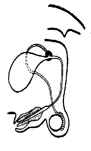
1. Add the following labels to the diagram of the reproductive system of a male dog shown below.
- vas deferens, urethra, testes, penis, scrotal sac, bladder, epididymis, penis bone, glans penis, prostate gland.
2. Fill in the table using the choices in the list below.
- A. Accessory glands; B. Epididymis; C. Vas deferens (sperm duct); D. Penis; E. Seminiferous tubules; F. Urethra
| Structure | Description |
| ......................... | 1. Organ that delivers semen to the female reproductive tract |
| .......................... | 2. Where sperm are produced |
| .......................... | 3. The tube that carries sperm from the epididymis to the urethra. |
| ............................ | 4. The tube that carries both sperm and urine down the penis. |
| ............................ | 5. Organs that contribute 90% of the semen. |
| ............................ | 6. Tubules where sperm are stored. |
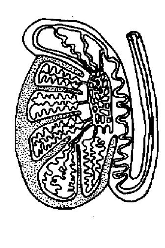
3. The diagram below shows a section through a testis.
Colour and label the structures in the diagram.
- 1. Seminiferous tubules in which the sperm are made. Blue
- 2. Collecting ducts where the sperm are stored. Green
- 3. Epididymis in which sperm mature and become motile. Red
- 4. Fibrous coat surrounding and protecting the testis. Brown
- 5. Vas deferens or sperm duct. Yellow
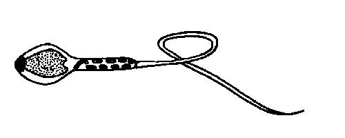
4. The diagram below shows a sperm. Colour and label the following areas.
- a) The DNA-containing area. Brown
- b) The enzyme-containing sac that aids sperm penetration of the egg. Yellow
- c) The midpiece - contains mitochondria for energy for sperm movement. Red
- d) The tail – propels the sperm along the female tract. Blue
5. a) What is the difference between sperm and semen?.........................................
- b) What is the difference between infertility and impotence?....................................
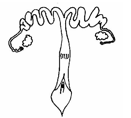
6. Add the following labels to the diagram of the female reproductive system below.
- ovary, vulva, fallopian tube, cervix, vagina, uterus
7. Fill in the following table with the words from the list below. (You may need to use some words more than once).
- A. ovary, B. vulva, C. fallopian tube, D. cervix, E. vagina, F. uterus
| Term | Description |
|---|---|
| ................................ | 1. Chamber that houses the developing foetus |
| ................................ | 2. Canal that receives the penis during copulation |
| ................................ | 3. Usual site of fertilisation |
| ................................ | 4. Duct through which the ovum travels to reach the uterus. |
| ................................. | 5. A sphincter muscle between the uterus and the vagina |
| ................................. | 6. External genitalia |
| .................................. | 7. Where the ova are produced |
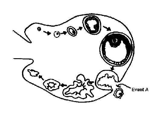
8. The diagram below shows an ovary with the stages of development of the ovum during an ovarian cycle.
i) Chose different colours and colour in:
- a) The cells that produce oestrogen.
- b) The structure that produces progesterone.
- c) All the ova.
ii) In the space provided, give the name of "event A’ on the diagram.
9. a) Arrange the following events in the ovarian cycle in the order in which they occur. Put the numbers in the correct order in the boxes below.
- 1. Luteinising hormone secreted by the anterior pituitary gland
- 2. Ovulation of mature ovum
- 3. Progesterone secreted by corpus luteum
- 4. Follicle stimulating hormone (FSH) secreted by the anterior pituitary gland
- 5. Corpus luteum develops
- 6. Ovum develops in the follicle
- 7. Oestrogen secreted by follicle cells
| ... | ... | ... | ... | ... | ... | ... |
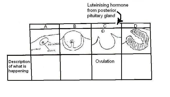
10 The diagrams below show different stages in the ovarian cycle.
- i. In the spaces under the diagrams write a few words describing what is happening in the diagram above. The third one has been done for you.
- ii. Now show by means of arrows added to the diagram, where the hormones FSH (follicle stimulating hormone), LH (luteinising hormone), oestrogen and progesterone act or are produced.
11. State whether the following statements are true or false. If false write in the correct answer.
- 1. The mixing of foetal and maternal blood in the placenta allows easy transfer of nutrients and oxygen to the foetus. T / F
- 2. Adrenaline cannot easily cross the placenta. T / F
- 3. Antibodies cannot pass across the placenta from the mother. T / F
- 4. Colostrum contains lots of hormones. T / F
- 5. Oestrogen stimulates milk “let down”. T / F
- 6. Young animals often have to be given iron supplements because milk contains very little iron. T / F
12. Insert the correct term into the table using the words below.
- A. progesterone, B. oestrogen, C. luteinising hormone, D. follicle stimulating hormone
- E. chorionic gonadotrophin, F. morula, G. blastocyst, H. implantation, I. placenta, J. colostrum
| Term | Description |
|---|---|
| ......................... | 1. The hormone that stimulates the growth of ovarian follicles. |
| ......................... | 2. The hormone that is secreted by the corpus luteum |
| ......................... | 3. A ball of cells produced by early division of the fertilised egg. |
| ......................... | 4. The hollow ball of cells produced by later division of the fertilised egg. Embryo transfer is possible at this stage. |
| ......................... | 5. The hormone that changes the empty follicle into the corpus luteum. |
| ......................... | 6. The membranes that form around the embryo to allow diffusion of nutrients and oxygen etc. between the foetal and maternal blood systems. |
| ............................ | 7. Attachment of the fertilized egg to the uterine lining |
| ............................ | 8. The hormone that is used in some pregnancy tests. |
| ............................ | 9. The hormone secreted by the ovarian follicle. |
| ............................. | 10. The first milk. |
