Fardeen khan Biology notes
Contents
O Level topics: Transport in Animal: Heart structure
Heart structure
Before we start this lesson we shall start with an activity to review what we know about Transport in animals
learners should be able to:
|
Introduction
The Heart – a muscular organ which drives the blood round the whole body Arteries - the blood vessels which carry the blood away from the heart. Veins – the blood vessels which convey blood towards the heart Capillaries – the microscopic thin walled blood vessels which carry blood from a small artery (arteriole) to a small vein (venule)
The heart: structure and function
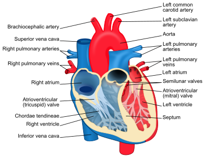
The upper thin-walled chambers are the atria (sing. Atrium) and each of these opens into a thick walled chamber the ventricle below. The wall of left ventricle is considerably thicker than that of the right ventricle. This is because the left ventricle has to pump blood round the body (except the lungs) whereas the right ventricle pumps blood a much shorter distance to the lungs.
Deoxygenated blood passes from the vena cava into the right atrium then into the right ventricle and through the pulmonary artery to the lung. Oxygenated blood enters the left atrium from the pulmonary vein and it passes into the left ventricle and then into the dorsal aorta for distribution round the body.
Pulmonary vein brings oxygenated blood from the lungs into the left atrium. The vena cava brings deoxygenated blood from the body tissues into the right atrium. The blood passes from each atrium to its corresponding ventricle and the ventricle pumps it out into the arteries. The aorta carry oxygenated blood to the body from the left ventricle. The pulmonary artery carries deoxygenated blood from the right ventricle to the lungs.
When pumping blood the muscle in the walls of the atria and ventricles contracts and relaxes. The walls of the atria contract first and force blood into the two ventricles. Then the ventricles contract and send blood into the arteries. Presence of valves prevent backflow of blood. Tricuspid valve (3 flaps) – between right atrium and right ventricle Bicuspid valve (2 flaps) – between left atrium and left ventricle. The flaps of these valves are shaped like parachutes with strings called tendons or cords to prevent their being turned inside out.
Semi-lunar valves (half moon) found in pulmonary artery and aorta. Each valve consist of 3 pockets which are pushed flat against the artery walls when blood flows one way. If blood flow back the pockets fill up and meet in the middle to stop flow of blood.
When the ventricles contract, blood pressure closes the bicuspid valves and tricuspid valves and prevent blood returning to the atria. When the ventricles relax, the blood pressure in the arteries closes the semi lunar valves so preventing the return of blood to the ventricles.
One complete contraction (systole) and relaxation (diastole) is called a heart beat. It takes less than one second (average heart beat = 70 times a minute)

