Microbial World

|
Unit 6: Microbial World |
By Dr. Abdul Karim
- Introduction
- Learning Objectives
- Bacteria
- Structure of the bacterial cell
- Bacterial reproduction
- Economic importance of bacteria
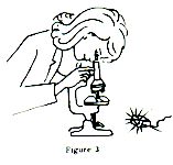
- Activity:Observasion of bacteria under microscope
- Virus
- Bacteriophages
- General morphology
- Multiplication of bacteriophages
- Economic importance
- Activity:Observasion of viral plant diseases
- Fungi
- Agaricus
- Economic importance
- Activity:Observation of yeast under microscope
- Let's Sum Up
- Key points
- Glossary
- Practice test
- Answers to SAQs
- References and further readings

|
Introduction |
You have learnt regarding organisms that can be seen with naked eye from the previous units of your learning material. Now we will discuss about organisms which can’t see with naked eye. It is so surprising, right?
We can see different types of plants are present both of water and terrestrial habitat. Among them some are too small that we can’t see them with the naked eye. These are the members of the kingdoms Eubacteria and Archaebacteria, are the most successful of all creatures on Earth, if success is measured by numbers of individuals.
Microorganisms are the living organisms of microscopic size, which include bacteria, fungi, algae, protozoa, and the infectious agents at the borderline of life that are called virus.In this unit, We will discuss elaborately about bacteria, virus and fungi .
| Bacteria |
Bacteria are generally chlorophyllless, prokaryotic (without nucleus), single celled microscopic (can’t see with the naked eye) organisms. The meaning if the word bacteria is stalk. In essence they consist of cell protoplasm contained within a retaining structure or cell envelope. They are among the most common and ubiquitous organisms found on earth. For example, even a gram of soil can contain up to 1010 bacteria. They can also be found in diverse and extreme environments.
| Structure of the bacterial cell |
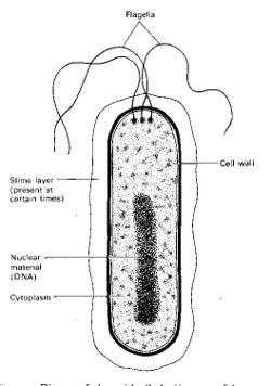
Different kinds of bacteria have different structures. Here we will discuss structure of a typical bacterial cell.
- Flagella A hair like appendages come out from the inner side of bacterial cell is called flagella (singular flagellum). Bacteria can move into the liquid medium with the help of flagella.
- Capsule A rigid layer of protein that is bounded to the cell wall and known as a capsule.
- Cell wall All bacteria are protected from the external environment by a layer called cell wall. It contains 10-40% weight of total bacterial cell.
- Cytoplasmic membrane It consists inside the bacterial cell wall. Sometimes, a pocket like small appendage developed inside the cell wall known as mesosome.
- Cytoplasm It covers with cytoplasmic membrane. Ribosomes and vacuoles are irregularly distributed within the bacterial cytoplasm.
- Nucleus Well-developed nucleus is absent in bacteria. But nucleoid is present almost centre portion of the cell. Nucleoid is made by a double stand DNA. Bacteria are prokaryotic due to absence of nuclear membrane and nucleolus.
| Bacterial reproduction |
Bacteria are multiplies through binary fission. It’s an asexual reproduction and sometimes called vegetative reproduction due to absence of any spore.
- Binary fission
Binary fission is the typical and main way to multiply bacteria. It is very simple and very fast reproductive method.
i) Cytoplasmic membrane of the bacterial cell developed into the inner side at middle of the cell and simultaneously nucleoid materials are also start to divide. Cell is divided into two cells after attachment of cytoplasmic membrane. Both of two daughter cells receive nuclear materials.
ii) Two new cells become mature.
iii) Newly developed cells are separated from each other and two bacteria are developed from a single cell.
Binary fission is occurred in a favourable condition and it takes about 20 to 30 minutes.
| Economic importance of bacteria |
Economically bacteria are very much important. Both beneficial and harmful effects are occurred in bacteria.
- Beneficial effects
i) Production of antibiotic Bacteria can produce some kind of antibiotic, as such-
| Bacteria | Antibiotic |
|---|---|
| Bacillus subtilis | Subtilin |
| Bacillus polymyxa | Polymixin |
ii) Preparation of beverage: Bacteria keep a vital role for the preparation of tea, coffee and tobacco.
iii) Leather industry: Bacteria help to remove the hairs from the leather.
iv) Remove of texture from jute: bacteria keep an important rope to separate texture form jute and there is no alternative to do so.
v) Vitamin preparation: Some bacteria like E. coli, Acetobacter acrogenes can produce different vitamins, as such Vitamin B, Thiamin, Riboflabin, Vitamin K etc.
vi) Decomposition: Bacteria decompose all death parts of plant and animal.
vii) Digestion of cellulose: Bacteria help to digest cellulolytic materials those were eaten by cattle, cows and goats.
viii) Production of milk product: Yogurt, Cheese, butter etc produced from milk after fermentation by bacteria.
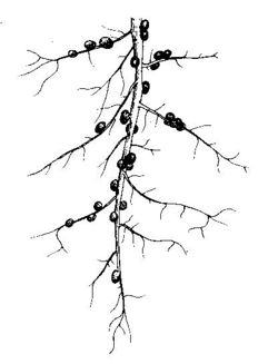
ix) Nitrogen fixation: Some soil bacteria like Azotobacter, Clostridium, Pseudomonas etc can fix nitrogen directly from the air and help to develop fertility of soil. Symbiotic bacteria also can fix nitrogen directly from the air.
- Harmful effects
i) Produce disease
a. Man: Bacteria can produce different diseases in human bodies. Such as-
| Disease | Bacteria |
|---|---|
| Cholera | Vibrio cholerae |
| Typhoid | Salmonella typhi |
| Tuberculosis | Mycobacterium tuberculosis |
| Dysentery | Bacillus dysenteri |
| Tetanus | Clostridium tetani |
b. Animals: Bacteria can cause the different diseases in household animals.
| Disease | Bacteria |
|---|---|
| Tuberculosis of cows | Mycobacterium bovis |
| Cholera of household birds | Bacillus anticepticus |
c. Plants: Bacteria can produce different disease in cultivated crops.
| Plant | Disease | Bacteria |
|---|---|---|
| Rice | Leaf blight | Xanthomonas oryzae |
| Wheat | Tundu | Agrobacterium tritici |
| Potato | Brown rot | Pseudomonas solanacearum |
d. Water Pollution: bacteria can causes the pollution of water and different diseases are spread through polluted water. Water born diseases like Cholera, Typhoid, Dysentery, Diarrhea etc occurs due to drink of polluted water.
| Virus |
Viruses are infectious agents so small that they can only be seen at magnifications provided by the electron microscope. They are 10 to 100 times smaller than most bacteria, with an approximate size range of 20 to 300 nm.
Viruses are incapable of independent growth in artificial media. They can grow only in animal or plant cells or in microorganisms. They reproduce in these cells by replication ( a process in which many copies or replicas are made of each viral components and then assembled to produce progeny virus). Thus viruses are referred to as obligate intracellular parasites.
A virion is a complete, fully developed viral particle composed of nucleic acid and surrounded by a protein coat. This coat protects it from the environment and serves as a vehicle of transmission from one host cell to another. Viruses are classified by differences in the structures of these coats.
Capsid A protein coat called the capsid surrounded the nucleic acid of a virus. The structure of the capsid is ultimately determined by the viral nucleic acid and accounts for most of the mass of a virus, especially of small one.
Nucleic acid
Virus contains a single kind of nucleic acid, either DNA or RNA, which is the genetic material. The percentage of nucleic acid in relation to protein is about 1% for the influenza virus and about 50% for certain bacteriophages.
In contrast to prokaryotic and eukaryotic cells, in which DNA is always the primary genetic material (and RNA plays an auxiliary role), a virus can have either DNA or RNA but never both. Generally plant virus contains RNA and animal virus contains DNA.
| Bacteriophages |
Federick W. Twort in England discovered bacteriophages, viruses that infect bacteria, independently in 1915 and by Felix d’Herelle at the Paseur Institute in Paris in 1917.
We will discuss here about T2-bacteriophage.
T2-Bacteriophage
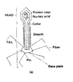
The common and well-known bacteriophage is T2. T2 bacteriophage is divided into two parts, are Head and Tail.
- Head The head of T2 bacteriophage is hexagonal. A double stand DNA is situated spirally inside the cell and a protein coat surrounded the nucleic acid.
- Tail A long tail is situated after head. Upper part of tail, such the connecting point of head and tail is called colar. The protein coat of tail is swollen and covered by protein. Six tail fibers and a baseplate present at the end of tail.
| General morphology |
Viruses may be classified into several morphological types on the vasis of their capsid architecture. The structure of these capsids has been revealed by electron microscopy.
Helical viruses
Helical viruses resemble long rods that may be rigid or flexible. The viral nucleic acid is found within a hollow, cylindrical capsid that has a helical structure.
Polyhedral viruses
Many animal, plant and bacterial viruses are polyhedral viruses; that is, they are many shaped. The capsid of most polyhedral viruses is in the shape of an icosahedron, a regular polyhedron with 20 triangular faces and 12 corners.
| Multiplication of bacteriophages |
Although the means by which a virus enters and exits a host cell may vary, the basic mechanism of viral multiplication is similar for all viruses. The best understood viral life cycles are those of the bacteriophages.
Phages can multiply by two alternative mechanisms: the lytic cycle or the lysogenic cycle. The lytic cycle ends with the lysis and death of the host cell, whereas the host cell remains alive in the lysogenic cycle.
| Economic importance |
Viruses are responsible to produce different kind of diseases in plants and animals. But viruses also play a positive role for human beings.
- Harmful effect
Virus produces different diseases to plants, animals and humans, as such-
i) human diseases like pox, hum, polio, influenza are produced by viruses.
ii) viruses are responsible for AIDS, a deathful disease of human beings.
iii) pox of cows; foot and mouth disease of cow, sheep, goat, and buffalo produce by virus.
iv) different mosaic diseases of plant, like leafroll, bushystant, tungro disease of rice etc approximately 300 kind of diseases are produced by viruses.
- Beneficial effect
Virus can play positive role to human being, as such-
i) different vaccines, like small pox, jaundice etc vaccines are produced from viruses.
ii) Viruses are used to scientific study in Molecular biology and genetic engineering.
| Fungi |
Fungi are eukariotic organisms. They can’t produce their own food because they have no chlorophyll. Thus, they either depend on a living host organism (in which case they are said to be parasites) or on decaying matter for their nutrition (thus called saprophytes). Like bacteria, their cell walls are made up of cellulose and chitin. There are many kinds of fungi. We may have seen them as green, white, gray, black, and orange molds on various things left in warm and moist conditions. The toadstools in the woods are another kind.
Mushroom, bread mold, and yeasts also belong to the group of organisms called fungi.
Fungi are widespread in the environment. Most fungi multiply asexually by forming many spores. These spores are easily carried to places by wind, water, animals, or humans. Some sensitive people develop allergy when they inhale these spores.
Fungi also multiply by budding. Other groups of fungi, however, reproduce sexually by fusing male and female hyphae in a process known as conjugation.
Hyphae are branching filaments consisting of several cells. They represent the vegetative or asexual stage in the life of most fungi. Along the hyphae, root like structures that attach the fungi to a surface extend downward. Hyphae absorb nutrients from that surface.
At certain points, structures that form spores extend upward. Spores are located in the sporangium. Black spots seen on stable bread are actually spores. These fruiting structures are used to classify fungi.
| Agaricus |
Agaricus is a saprophytic fungus. Species of these types of fungi are called gill. Agaricus can grow on muddy soil, upper side of wood, compost manure, rotten straw, Cow dung etc. Agaricus can grow well in rainy season. Sometimes it can grow as a circle within the grass field. This type of cycle is called pericycle.
Structure
The somatic structure of agaricus divides in two parts, one is vegetative part or mycellium and another part is reproductive part or fruiting body.
Vegetative mycellium of agaricus develops from the primary mycellium of germinating basidiospore. Mycellium have many branch and sub-branches, and grow within the moist soil. Haphae of mycellium are septate. Hyphae are white colour and take nutrients from habit.
Cells of hypha consist of many nucleous, grnular cytoplasm, small vacule and oily drops. Mycellium of hypha sometimes aggregates together and formed a rope like structure called rhisomorph.
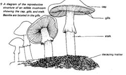
Agaricus consists of a stalk with an umbrella-shaped cap. On the cap’s undersurface are gills. Fruiting bodies called basidia that produce spores are found on the side of gills. Mature spores fall to the ground. They may be carried by wind or water to other area. Mature fruiting body looks like umbrella and that’s why it is called toad’s umbrella.
| Economic importance |
Mushrooms are another kind of fungi. Some mushroom species are edible and others are poisonous.
Nowadays, edible mushrooms such as Volvariella sp. are commonly grown. Ipil-ipil leaves, rice stalks, and banana leaves are used as growing surfaces. To these substrates, honey, coconut water, and urea may be added.
Life saving antibiotic can produce from different fungi. Penicillin produces from Penicillium notatum. Sir Alexander Fleeming was discovered Penicillin in 1928.
Some fungi are also decomposers like bacteria. They can also cause spoilage of food. Fungi decompose organic materials.
Some fungi are parasitic on humans, animals, and plants. In humans, they can cause ringworm, athlete’s foot, and other skin diseases. Certain fungi may be caused for Wilting of crops like tomato, corn, banana, and papaya. Fungi may also attack seeds. Consequently, these seeds don’t germinate.

|
Results |

|
Key Points |
The key points of this chapter are as follows:
- cellular
- bacteria
- fungi
- microscopic
- parasitic
- prokaryotic
- saprophytic
- virus

|
Glossary |
cellular. with cellular structure
microscopic. can’t see with naked eye
parasitic. live on a host plant or animal from which take nutritious material
prokaryotic. without typical nucleous
saprophytic. live on dead materials

|
Practice Test |
- Short Answered Questions
- What is bacteria?
- Which bacteria can produce polymyxin?
- What is fungi?
- Name a common fungi?
- Virus is living or no-living organism?
- What is the chemical composition of virus?
- Essay Type Questions
- Describe bacterial structure and it's multiplication processes.
- Explain bacterial economic importance.
- Draw and describe somatic feature of Agaricus.
- Describe somatic structure of virus.
- Discuss economic importance of virus.

|
Answers to SAQs |
SAQ-1
A. Stalk
B. Binnary fission
C. Bacillus subtilis
SAQ-2
A. Acellular
B. Protein and nucleic acid
C. RNA
D. DNA
SAQ-3
A. Saprophytic
B. Agaricus
C. 1928
D. Penicillium

|
References and Further Readings |
- Karim, M.A., S.A. Sultana, M.A.Islam, M.S. Islam and A.H.M. shamim. 2006. General Science. CAMPE and Bangladesh Open University.
- Hoque, M.E., M.A. Karim, S.A. Sultana and S. Sultana. 2003. General Science. Bangladesh Open University.
- Fattah, Q.A., I.A. Muttaki, Faridunnesa, A.K.M.N. Islam, K. Banu, M.A. Hasan and M.A. Karim. 2004. Biology-I. Bangladesh Open University.
- Pelczer, M. J., E.C.S. Chan and N.R. Krieg. 1986. Microbiology (5th Ed.). Tata McGraw-Hill Publishing Company Ltd. New Delhi.
- Purves, W.K., G.H. Orians and H.C. Heller. 1995. Life: The Science of Biology (4th Ed.). Sinauer Associates Inc., Massachusetts, USA.
- Maier, R.N., I.L. Pepper and C.P. Gerba. 2000. Environmental Microbiology. Academic Press, UK.


