Microbial Animal Interactions
This is based on New Zealand Qualification Authority Unit 8022 [1] entitled "Demonstrate knowledge of microbial-animal interactions."
Contents
Element 1
The Bacterial Flora of Humans outlines the normal flora of humans
1.1
Micro-organisms are various microscopic organisms too small to be seen with the naked eye. They include mainly bacteria, but also fungi, viruses, yeasts etc. Approximately 90 trillion microbes continuously inhabit the human body!
A mixture or a group of organism that commonly found at any anatomical site is referred to as the normal flora. In a healthy animal blood, brain and muscle are normally not affected by normal flora but the surface tissues like skin and mucous are constantly exposed. Normal flora of humans is complex it varies according to age, sex, diet and nutrition. Microbial association with skin is mainly due to staphylococci and Corynebacteria. These two are considered as non pathogenic.It has some parasitic and mutalistic roles too, it produce fatty acid that inhibit the growth of fungi and yeast in the skin. In certain occasion it act as a pathogen especially humans who are nasal carriers, their face and hands are likely to be infected with bacteria. They cause infection themselves and spread to others.[2] [3] Some of them reside on skin, some in the respiratory system, while others inhabit the alimentary tract.
Alimentary tract: Found in multicellular organisms, this is an internal system or 'canal' consisting of; a mouth, esophagus, stomach, intestines, rectum and anus. Its function processes food by ingestion, digestion, extraction of nutrients and finally, elimination of waste materials from the body.[4]
The surface of skin is not a favorable place for microbial growth because it is often dry, salty, and has low pH. Most microorganisms are associated with sweat glands and hair follicles because of the moist and nutritious environment. Urea, amino acids, salts, lactic acids, and lipids are secreted through the skin and provide microorganisms with what they need to grow[5].
The upper respiratory tract in a human is an ideal habitat for aerobic micro organisms. There is plenty of oxygen, moisture, nutrients available and mucus to adhere to. “Normal Flora” in the upper respiratory tract can include Actinomyces sp., Candida sp., diphtheroids, Haemophilus sp., Neisseria sp., Staphylococcus sp. and Streptococcus sp. Most of these organisms have a relationship with a person that is mainly beneficial to the microbe which is called commensalism. The one main benefit they do provide for the human is that they keep out other pathogens by colonizing all available space. However, if the person’s immune system is compromised, then these microbes can cause serious infection.
1.2
Micro-organisms found in the alimentary tract may have either good or bad effects on the host animal. The disadvantages may be conditions like gastroenteritis or pathogenic disorders like poisoning of food by bacteria such as Salmonella. Benefits of course are interactions involved in digestion of food within the stomach and intestines. An example here being bacteria like Lactobacillus bifidus and Lactobacillus acidophilus which aids the digestion of lactose.]] Assimilation of vitamins such as Vitamin K, essential for blood clotting is also aided by micro-organisms. Animal bodies then, provide the ultimate habitat for micro organisms to survive. With adequate nutrient availability, moisture, pH and temperature levels maintained, environmental conditions are optimal for survival.[6]
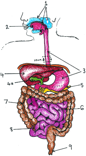
Components of the Mammalian Alimentary Canal
1-Salivary glands- first of the accessory glands. Consisting of three pairs of glands that secrete digestive enzymes. In saliva the enzyme salivary amylase, hydrolyses starch from plant materials and glycogen from animal matter. Generally 1010 per ml bacteria exist here. Streptococcus salivarus is consistently extracted from infants, even as early as two days after birth. Saliva contains buffers and bactericidal agents that render most bacteria useless. The majority of bacteria present maintain a 'normal flora'.
2-Tongue- an agile organ that manipulates food around the mouth while chewing and helps form a bolus (food ball). It moves the bolus to the back of the oral cavity and ensures it is at the entrance to the esophagus.
3-Esophagus and Stomach- involuntary muscle action (peristalsis), moves the bolus toward the stomach, taking 5-10 seconds. Preliminary digestion takes around 2-6 hours with sphincter muscles preventing food escaping until necessary. The lining of the stomach wall secretes gastric juice. Pepsin, the enzyme here, withstands the highly acidic (pH 2) conditions and disrupts the matrix binding animal and plant cells, breaking them into smaller peptides. Only extreme acidophiles like Heliobacter pylori can exist-until treatment with antibiotics eliminates them. Micro-organisms of less than 10 per ml generally survive.
4-Liver 4a-Gall bladder 5-Pancreas
All are accessory glands responsible for secretion of, or storage of, enzymes aiding digestion of various foods according to the systems requirements. Protein, nucleic acid and fat digestions are aided by these organs. Hydrolisation is related to acid chyme, the continual churning of stomach contents, and release into the duodenum - first 25cm of small intestine.
6-Small Intestine- as long as 6m in adults it functions mainly to absorb nutrients and water from food as it travels by peristalsis. Of the micro-organisms present (usually 103 exist) most are gram + cocci and bacilli. In the ileum (end part of the small intestine) bacteria present resemble those of the large intestine, anaerobic bacteria and members of the Enterobacteriaceae family.
7-Colon (Large Intestine) - has the largest microbial population. Over 300 different species have been isolated from human faeces, and make up 50-60% of its total dry weight. Health, food type and water content effect the microbial make up of the colon. Facultative anaerobes present belong to the genera Escherichia, Proteus, Klebsiella and Enterobacter. Escherichia are the more prevalent and have provided molecular biologists with the most entertainment and/or research material. Travel along the colon takes about 12 to 24 hours as almost 90% of ingested water is removed.
8-Appendix (Cecum in other organisms) - removes any foreign objects that escape digestion. Contains no differing micro-organisms but irritation by bigger particles may cause inflammation. Lymphoid tissue may make minor defense contributions.
9-Rectum- Terminal portion of the digestive canal where faeces is stored prior to elimination via the anus. Contents (faeces) has a very high microbial count. E. coli present here in high numbers, is, in water supplies an indicator of faecal contamination! (see details in Water Treatment).
Skin Flora
Some of the most commonly found microorganisms on the skin are Corneybacterium diptheriae, Staphylococcus aureus, Micrococcus luteus, Staphylococcus epidermis, and Pityrosporum ovale. Most bacteria are beneficial to the skin because they prevent colonization of the skin by pathogens and they control the other organisms on the skin. But if a cut is present, these bacteria can enter the body and cause damage.
The following link leads you to brief descriptions of these common microorganisms and the diseases they can cause.
Microbial Flora of Skin
[Power Point presentation - Microbes: Animal Skin][7]
Element 3
Microbiology of the rumen consists of bacteria, fungus and protozoa. Bacteria can be grouped according to their shape, size and structure and also they are grouped eight according the type of substance fermented, they contribute 1010-1011gm/cells of the rumen. These groups utilize cellulose, hemicellulose, starch, sugars, immediate acids, protein and lipids to produce methane. These groups of bacteria are liable for all fermentation process in rumen; they remove hydrogen by the reduction of carbon dioxide to produce methane which helps methionic bacteria to increase the growth of other bacterial colonies. Protozoa is about 105 to 106 cells/gram of rumen contents and fungi contribute 8%, anaerobic fungi are the predominant one.
Protozoa act as a source of protein by ingest bacteria and also act as a stabilizing factor for fermentation end products. Bacteria and fungi helps in fiber digestion and anaerobic fungi degrading cellulose and xylene, to indicate their role in fiber digestion.
Microbes located in rumen mainly cover three phases, Liquid phase: occurs free living organism present in rumen fluid which feed carbohydrate and protein, it contributes about 25%.
Solid phase: Microbial group attached with food particles helps to digest insoluble polysaccharides like starch, fiber and less soluble proteins. It contributes 25% of microbial mass.
Last phase: 5% of microorganisms either attached to epithelial cells in the rumen or to protozoa.[8]
3.1
Ruminant Digestion
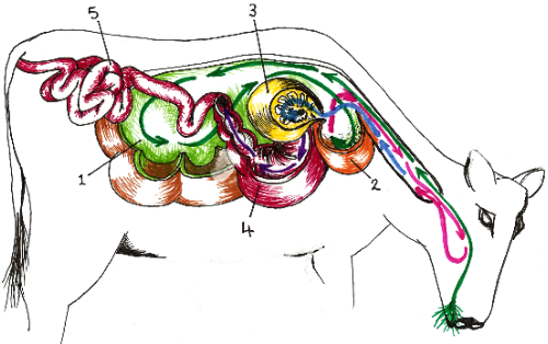
Key to Picture
1. Grass boluses (food balls), move from mouth, down the o esophagus, to the rumen. Rumen is a large fermentation chamber (approximately 125 litres in adult cattle), with textured surface and lined with papillae. Large number of micro-organisms survive in rumen mainly bacteria which secretes enzymes for cellulose digestion. Internal pH, temperature, and microbial population (1010 per ml) soften and ferment the cellulose-rich diet of grass.[9]
2. Food enters the reticulum where further symbiotic prokaryotes and ciliated protista further digest the cellulose fibers. Due to the continuous build-up of internal gases from microbial action, ruminants belch and regurgitate their 'cud'.
3. Cud is chewed and swallowed again to enter the omasum. A high surface area provided by this organ removes most remaining water.
4. Fermented cellulose now enters the abomasum, the ruminants fourth 'stomach'. The cattle's own enzymes now digest and absorb remaining nutrients. Thanks to microbial action, ruminants absorb more nutrients than grass actually contains.
Carbohydrates digestion: These are digested by rumen microbes are converted into volatile fatty acids which are further absorbed in blood streams and are transported to all body tissues for its growth, maintenance and reproduction.
Protein digestion: These are broken down to ammonia which bacteria require for synthesizing their own body protein.
Rest of the ammonia is absorbed in blood streams, carried to liver, converted to urea and excreted in the form of urine. Remaining urea is returned to the rumen via saliva and directly through the rumen.
Microbes in rumen manufacture all B and K vitamins.[10]
There are three main groups of microbes in rumen of cow:
Bacteria digest of sugars, starch, fiber and protein for cow.
Protozoa swallow and digest bacteria, starch granules, and some fiber.
Fungi is a minor group but do important job by splitting open plant fibers to make them more easily digested by the bacteria[11].
For more information on the rumen and its digestion, plus other facts on the rumen see: http://en.wikipedia.org/wiki/Rumen
Rumen Microbes and Nutrient Management outlines the major microbial groups and their metabolic activities which contribute to digestion in the rumen.
[Power Point presentation - Ruminant Animals][12]
Element 4
The web site Animal Disease Information provides a broad range of animal disease with image, factsheet and diagnosis. And some other useful information as well.
4.1
Microbial pathogen can grow in any place which provides them with suitable environment for growth. For example soil, air, insects or animal body etc it rapidly grows and multiplies so that it can be transmitted to a susceptible animal. Animal disease can be classified as infectious and non infectious. Infectious caused by an agent (bacteria, virus, parasite or fungi).
Non infectious caused by factors like diet, environment, injury or heredity (some times the causes are unknown). Ref: microbiology principles and applications (second edition) By Jacquelin G Black.
Animal disease refers the disorder that affect animal health and ability to function, it reduces the productivity of animals that seriously affect economy of many industries, and some of them affect humans too. To identify a disease a veterinarian must consider the age, sex, breed and species of animals, because some diseases are prevalent in certain categories.
Bacterial disease Avian Tuberculosis
Animal infected: Birds
Source: Respiratory aerosols
Anthrax
Animal infected: Dogs, cats and domestic animals
Source: Contact with animals, contaminated soils, and hides and also ingestion of contaminated milk or meat
Salmenellosis
Animal infected: Dogs, cats, poultry and rats
Source: Ingestion of infected tissues and contaminated water
Brucellosis
Infected animal: Domestic animals
Source: Direct contact with infected tissues and ingestion of milk from infected animal
Leptospirosis
Infected animal: dogs, rodents
Source: Contact with urine, infected tissue and contaminated water
Viral disease Rabies
Infected animal: Dogs, cats, bats and wolves
Source: Bites, infected saliva in wounds
Encepalitis
Animal infected: Horse, birds and domestic animals
Source: Mosquitoes
Lassa fever
Infected animal: Rodents
Source: Urine
Parasitic & fungal diseases
Histoplasmosis: (fungal): Infected animal: Birds Source: Aerosols of dried infected faeces
Ring worm (fungal) Animal infected: Cat, dog and other domestic animal Source: Direct contact
African sleeping sickness (parasitic): Animal infected: Wild game animals Source: Tsetse flies
Toxoplasmosis (parasitic): Animal infected: Cats, birds, rodents, and domestic animals Source: Aerosols, contaminated food and water, placental transfer
Tape worms (parasitic): Animal infected: cattle & rodents. Source: Ingestion of cysts in meat
Ref: microbiology principles and applications (second edition) By Jacquelin G Black
There are many places where a person can pick up pathogens.
Person to Person Contamination:
Coughing and sneezing passes on viruses like influenza by aerosols that are breathed in by another person. Bacteria like Mycobacterium tuberculosis is also passed on this way.
Faecal-Oral is a common way to pass on Hepatitis B and many food pathogens. A person infected with Hepatitis B may go to the toilet and not wash their hands thoroughly. This person may then prepare food for another person. This next person eats the contaminated food and contracts Hepatitis B.
Re-used equipment or equipment not properly sterilised has been proved to be a serious problem for pathogen control. Beauty Therapy and Tattoo outlets have been known to pass infections (Hepatitis C) through re-using needles and other equipment, such as Staphylococcus aureus which can cause Toxic Shock Syndrome. Contaminated needles can also pass on HIV.
Bodily Fluids spread many diseases easily. Sexually transmitted diseases included HIV, Chlamydia and Syphilis. These are becoming increasingly common and can have serious consequences such as infertility.
Environmental Contamination:
Water can contain protozoa like Giardia and Cryptosporidium. These microbes are picked up when contaminated water is drunk. They cause abdominal pain and diarrhea. Faecal contaminated water is a large problem in developing countries. The water can carry life threatening pathogens like Vibrio bacteria (cholera and other vibrio illnesses), Shigella and Salmonella bacteria (dysentery and typhoid).
Soil harbours many pathogens as it is a habitat for a large range of microbes. Animals easily come into contact with these soil-borne pathogens because most of our food source comes from soil. Vegetables, fruit, wheat all grow in the soil and farm animals live and ingest any microbes on the grass that they eat. These pathogens can be also passed down the food chain, e.g. a cow eats contaminated grass, pathogen replicates inside the cow. The cow is killed for meat, which a human ingests and is infected with the pathogen. Water also filters through the soil, picking up any pathogens along the way and contaminating waterways. As the soil dries to dust and is stirred up, aerosols are created. The aerosols are breathed in and infection can then start in the lungs. Examples of soil pathogens are Clostridium perfringens (gas gangrene), Clostridium tetani (tetanus) and Bacillus antracis (anthrax).
Sources, Types and Pathways identifies sources and reservoirs of major microbial pathogens of animals.
4.2
Salmonellosis is a bacterial infection caused by salmonella. Symptoms associated with diarrhea and septicemia, it is one of the main diseases fond in birds, reptiles, wild and domestic animals. Route of entry is mainly through mouth commonly by food and water contaminated with infected faeces or by direct contact with infected animal and multiplies in small intestine. And also it transmitted by object contaminated with infected animal products like eggs or [13]. Certain factors are there to increase the spread of this disease, increase in animal husbandry and production are the main reasons. Due to the increased number of farm animals' protein and vegetable by products imported in a large scale, which result in the widespread of disease in animals and humans.
Pathogenesis of Foot and Mouth disease: It is usually transmitted by aerosol, with the infectious particles which are exhaled by infected animal. The infectious particles are carried by air to the respiratory tract of susceptible animal. The incubation period to observable disease is 2 to 8 days. Further the virus goes under the process of replication. Vesicles develop as the virus grows within the group of contiguous epithelial cells, rupturing them and creating a lake of fluid within the epithelium. The vesicular fluid contains abundant viral particles and the virus persists in the surrounding cells for 3 to 8 days.[14]
Fleece rot and dermatophilosis in sheep outlines the process of pathogenesis of Pseudomonas aeruginosa on sheep.
4.3
Many countries prevent salmenellosis in animals for preventing many food born diseases. Salmonella typhimurium DT104 is the anti bacterial drug used for controlling disease in animals. The feeding of faeces from adult hen to young is found to be a good method to prevent bacterial colonization, so it is used in poultry industry.[15]
Control for Foot and Mouth disease
Disinfection of infected materials.
Quarantine measures.
Destruction of infected products in infected area.
Infected animals should be kept away from other animals.
Proper cleaning should be done of the premises.
Fleece rot and dermatophilosis in sheep indentifies some control mechanisms for fleece rot.
[Power Point presentation - Disease Process in Animals: foot rot in cattle][16]
Element 5
Here is a lecturing material that I hope of helping you to understand Infectious Disease Epidemiology.
Epidemiology is the study of factors and mechanisms that present in the spread of disease within a population. Epidemiologists are the scientists who study epidemiology and causes and transmission of infectious disease. They classified diseases according to the size of geographic area affected and the degree of hazard the disease cause in population. On the basis of that disease can be classified as endemic, Epidemic and pandemic or sporadic Endemic disease is occurs constantly in a particular geographic area degree of severity and the number of cases are less and it vary according to the different geographic area and it is not consider as a public health problem. Mumps and chicken pox are the examples of endemic disease. Epidemic disease has a sudden and high incidence within a population, the severity and the number of cases is high and considered as a public health problem. It is caused by the presence of a particular virulent strain of a pathogen in a large population who lacks immunity. Polio myletics is considered as a epidemic disease and a severity of the epidemic disease is determined according to the pathogen and the type of host. Pandemic disease occurred when epidemic become world wide. Cholera is the most recent pandemic it spread 1961 to1971 in various part of the world.
There are few methods which used control the transmittable diseases.
Isolation: In which person having particular communicable disease is prevented from general population.
Quarantine: Separation of human or animal from common population having communicable disease have been exposed to one.
Immunization: Control of communicable disease by using safe vaccines.
Vector control: Is considered as an effective means of controlling infectious disease in which a vector or an organisms can be identified and also its habitat, feeding behaviour, breeding to be considered and then the place which lives as well as breeds should be destroyed by an insecticides and rodenticides.
Ref: microbiology principles and applications (second edition) By Jacquelin G Black
5.1
An infectious disease may result in outbreaks easily depending on the ease of micro organism that can move from one animal to another. An Outbreak may result into an Epidemics or Pandemic. An infectious disease which spreads beyond a local population in a wide area is called Epidemics. As soon as it becomes worldwide it is considered as Pandemic.[17]
An example of how the transmission of infectious diseases may result in localised outbreaks, epidemics and pandemics would be SARS (Severe Acute Respiratory Syndrome). The virus's first outbreak appears to have started in Guangdong, China. It then spread to Hong Kong and to other neighbouring countries, before spreading further by infected people flying to other parts of the world. See link http://en.wikipedia.org/wiki/Progress_of_the_SARS_outbreak
Epidemic outlines how the transmission of infectious diseases may result in localised epidemics.
5.2
Surveillance: It is an effective method of controlling infectious disease and preventing it from entering and causing outbreaks. It is a well ordered and timely response which is very effective to the outbreaks. It can easily detect re-emerging as well as newly developing infectious diseases.[18] Typing method: it is a method which helps in the investigation and detection of emergence of antimicrobial resistance and newly evolving pathogenic strains. Moreover it is useful in studying various other characters of a particular behaviour and other conditions which favour its growth, its developing time, mutations it can undergo etc.[19]
Meningitis caused by Meningococcal group B bacteria has been a recognised epidemic in New Zealand since 1991. During this time, it has caused 218 deaths, many of them being infants and young children. More than 5,400 have contracted the disease. Some have that have survived the disease have suffered permanent disfigurement and/or disability, such as loss of limbs and brain damage. The epidemic was expected to continue for at least another 10 years. So as a way to slow and stop the spread of the disease, The New Zealand Ministry of Health launched a immunisation campaign free for all New Zealanders under 20 years of age. See this link for more details on the immunisation program http://www.beehive.govt.nz/node/20259
For more information on Meningococcal B see link http://www.moh.govt.nz/moh.nsf/0/F9A713229EB450A3CC256C7B00734F5D/$File/1MeningococcalDisease.pdf
Are Active Microbiological Surveillance and Subsequent Isolation Needed to Prevent the Spread of Methicillin-Resistant Staphylococcus aureus outlines ways to control infectious diseases.
[Power Point presentation - Epidemiology: animal disease][20]

