Lesson 16: Oral, Eye and Skin Conditions
Contents
INTRODUCTION
Welcome to Unit 16 of our Child Health Course. In the last unit, you covered helminths or common parasitic worms that affect children. As you learned in that unit, the spread of helminths in children can be prevented. In this unit, we shall learn about common oral, eye and skin conditions that affect children. We shall also discuss common surgical conditions in children.
|
By the end of this unit you should be able to:
|
We shall start by discussing common oral health problems in children.
16:1 ORAL HEALTH
In this first section, we shall examine closely the causes of four common oral conditions in children. Common oral conditions, such as dental decay and gum infection, are widespread but can be prevented through simple daily good habits. We hope by the end of this section that you shall be able to diagnose, dispel myths and misconceptions, manage and prevent common oral conditions in children.
16.1.1: WHAT IS ORAL HEALTH
|
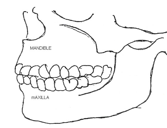
Oral health refers to the total wellbeing of the mouth. The word oral refers to the mouth. The mouth is one of the most important parts of the body. It is the first part through which the food we eat enters the body. The mouth comprises of the lips, teeth, tongue, cheek, palate, gums, and jaws (mandible and maxilla). See Fig. 16.1. Any of these tissues can suffer from infection. |
Fig. 16.1: The Structure of the Mouth
A human being has two sets of teeth. The teeth which erupt first are the primary teeth (also called deciduous teeth, temporary or milk teeth). The eruption of the primary teeth starts at around six months of age. Thereafter, a tooth errupts every month so that by the age of two and a half years a child has 20 teeth, the full set of primary dentition. The shedding of the primary dentition begins at about six years of age.
The second set of teeth is known as the permanent dentition, which replaces the primary teeth. The first permanent tooth errupts at about the age of six years. By the age of twenty-one years, most adults have thirty-two permanent teeth, the full set of permanent teeth.
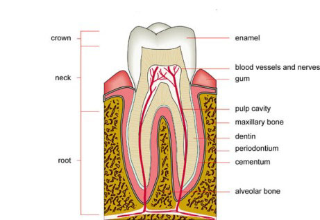
A tooth has the following parts as is shown in Figure 16.2:
- Crown: This is the visible part of the tooth above the gum.
- Root: The invisible part of the tooth below the gum.
- Enamel: This is the white part covering the crown, and is the hardest tissue of the body.
- Dentine: This is the main bony part of both the crown and the root. It is sensitive to pain.
- Pulp: This is the soft inner part of the tooth that contains the blood vessels and nerve endings. The pulp serves as the connection between the body and the tooth. It is the nutritional channel for the tooth.
Fig. 16.2: The Molar Tooth
There are 4 types of teeth depending on their function. These include:
- Incisors or front teeth: Used for cutting.
- Canines: The pointed teeth used for tearing.
- Pre-molars: The narrow teeth behind the canines used for chewing.
- Molars: The large teeth, larger than the pre-molars, used for chewing and grinding.
Teeth are necessary for feeding, speech, smile, and maintaining a good appearance.
| 1
What are the common dental problems among children in your area? _____________________________________________________________________ _____________________________________________________________________ _____________________________________________________________________ _____________________________________________________________________
|
16.1.2: COMMON ORAL PROBLEMS IN KENYA
The two most common and widespread oral conditions are tooth decay, also known as dental caries or cavities and gum infection, also known as gingivitis. The other two conditions we shall consider are malalignment and oral thrush.
Let us look at each in turn starting with dental caries.
Dental Caries:
Tooth decay is the slow destruction of the crown or root of the tooth by bacteria. Dental caries or tooth decay is one of the two common dental diseases that are responsible for most teeth loss in children.
The development of dental caries is a long process.
Causes of tooth decay The process starts when the bacteria in the plaque digest sugar, producing an acid. The acid, in turn, acts on the tooth surface dissolving a little bit of the tooth structure to form a small hole. The rate at which this hole increases depends on oral hygiene and the frequency of consuming sugary foodstuffs. This means that each time such foods are eaten, the decay increases.
You probably are seeing a white substance against your fingernail. This is what is referred to as PLAQUE.
A plaque is a sticky white or yellow deposit found on the teeth. Plaque provides a medium in which bacteria grow. There are always bacteria present in the mouth. Most of the bacteria living in our mouth are harmless and some even help in digesting food.
The dental decay process can be expressed by the following equations:
PLAQUE BACTERIA + SUGAR = ACID.
ACID + TOOTH + TIME = DECAY
The bacteria growing in the plaque ferment the carbohydrates to produce acids. The acids demineralize the tooth and so a cavity results.
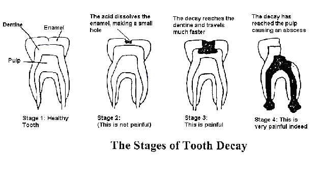
The diet we eat consists of a mixture of foods, and within one food there are many different substances. The group of substances responsible for dental decay includes refined carbohydrates such as chocolates, sweets, cakes, fizzy drinks, toffees, biscuits, sugar, etc. Fig. 16.1.3 below illustrates the stages of dental decay.
Fig. 16.3: The Stages of Tooth Decay
What are the signs and symptoms dental caries?
We shall discuss the signs and symptoms of each of the three different stages shown in figure 16.1.3.
In the first stage, there is a healthy tooth. In the 2nd stage, the dental decay is still in the enamel. The enamel, as we discussed earlier, has no living cells. Therefore, the initial stage of the tooth decay is not painful at all. In this stage, you should look out for visible changes in the tooth.
As the decay advances towards the dentine, the sensitivity can be felt especially with temperature changes in the mouth. In the 3rd stage, the decay involves the dentine, and the pain intensifies. The child complains of pain if they eat hot, cold, sweet and acid foodstuffs they complain.
The pain intensifies and becomes increasingly constant as the decay advances towards the pulp. In the 4th stage of decay, the pulp is reached. Remember that the pulp has both nerves and blood vessels so it is very sensitive.
So the 4th stage of dental caries is characterized by pain and a visible hole commonly referred to as a cavity. At first, the cavity may appear as a colour change on the tooth but in time it grows into a real cavity.
Untreated dental caries kills the tooth and then an abscess may form as illustrated. A dental abscess is a collection of pus around the root of the tooth with dental caries. A child with a tooth abscess will complain of a swollen cheek and a lot of pain. A fever may also be detected.
| 2
List the signs and symptoms of a child with tooth decay: _____________________________________________________________________ _____________________________________________________________________ _____________________________________________________________________ _____________________________________________________________________ Think about the children you see daily in your clinic. Describe the treatment you would give a child with a decayed tooth: _____________________________________________________________________ _____________________________________________________________________ _____________________________________________________________________ _____________________________________________________________________
|
Treatment
The choice of treatment for a decayed tooth depends on the severity and the location of the cavity. Treatment ranges from a simple filling of a cavity to tooth extraction. Proper dental treatment is available in established dental clinics. You should therefore refer such children for treatment. However, in case of a dental abscess, you should prescribe an antibiotic and pain killers and then refer for proper dental treatment.
Discourage the application of aspirin tablets on the affected tooth, as this will not treat the dental caries. It will cause an ulcer instead.
If a child loses a tooth due to dental caries, there are other forms of treatment to replace it. They include artificial teeth. Artificial teeth are expensive, however, and it is cheaper in the long run to take proper care of teeth and to have caries treated in good time.
Prevention:
Before we complete our discussion on dental caries, let us look at the various ways of preventing this problem. Start by doing the following activity.
| 3
How would you prevent the occurrence of dental caries in your community _____________________________________________________________________ _____________________________________________________________________ _____________________________________________________________________ _____________________________________________________________________
|
Now read through the text below and see if your ideas are included.
There are three main ways of preventing dental caries in children:
a. Decreasing the frequency of taking sugary foods prevents dental caries. Instead of taking sugary foods, children should be encouraged to eat alternative foods that are good for teeth. These include fruits, carrots, ripe bananas, sugar cane, and salty snacks such as groundnuts.
|
It is better to clean your teeth sufficiently, at least twice each day, especially after meals, than to clean four times unsatisfactorily
|
b. Oral Hygiene: Besides reducing the intake of sugary foods, thorough daily brushing and flossing will help in preventing dental caries. It is recommended to clean teeth with a toothbrush and tooth paste at least after every meal. Tooth cleaning can also be done using a tooth stick. Toothpaste with fluoride is useful because it cleans the teeth thoroughly and the fluoride content strengthens the teeth against decay. Parents should help children below 8 years with teeth brushing. The extent of the parental involvement should be according to the child’s ability.
c. Regular dental check-up: The third way of preventing dental decay is having a child’s teech checked regularly, at least twice in a year. This will enable early detection and treatment of any cavities. You can examine a child and if you identify a dental problem, give treatment or appropriate advice.
d. Using plastic dental sealants soon after the eruption of the teeth. A dental sealant is a plastic coating applied to the chewing surfaces of back teeth. In children, grooves in these teeth are so narrow that a toothbrush cannot fit into the spaces to remove plaque. Dental sealants prevent dental decay in the grooves. The sealant is applied on permanent molars as soon as they erupt.
e. Nutrition: Promoting healthy development of teeth and gums is another way of preventing dental caries. We have seen that diet can affect the teeth after they have erupted in the mouth but nutrition can also affect the development of teeth. It is important, therefore, for children to eat a diet rich in Calcium, Phosphate, and Vitamins C, A, D and protein in order to promote normal development of their teeth. Pregnant mothers should do the same to ensure proper development of their unborn child's teeth. Calcium and phosphate are found in milk and milk products. Vitamins are found in vegetables, protein is found in milk, meat, fish, eggs and legumes.
f. Optimizing the fluoride content of the water is the most effective preventive measure against dental caries.
Gum Infection or Gingivitis
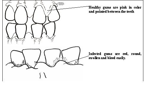
The second most common oral health problem in children is gum infection or gingivitis. The gum is also known as the gingivum and is the tissue and mucous membrane surrounding the tooth and alveolar bone as illustrated in Fig 16.1.4. Gingivitis is the slow and relatively painless inflammation of the gum.
Figure 16.4 : Difference between healthy and infected Gums
Causes of Gingivitis Like cavities, gum infection comes as a result of DENTAL PLAQUE. If the plaque is not regularly or properly removed from the teeth surfaces, it accumulates and hardens to form TARTAR or calculus. Tartar constitutes a suitable environment for the multiplication of bacteria. These bacteria continue producing acid. Calculus also makes teeth and gums cleaning very difficult.
Children Most Affected by Gingivitis
The prevalence of gingivitis increases with age. The age of onset is around 3 - 5 years. By puberty almost all children have gingivitis. This increase in the prevalence of gingivitis may be attributed to an increase in the gingival sites at risk as the permanent dentition develops. After puberty, there is a temporary decline in prevalence and severity. This decrease in prevalence and severity may be attributed to an increased social awareness, which results in improved oral hygiene. Besides this, some conditions such as malnutrition, poor health including mental disability, and certain habits, such as breathing through the mouth, increase chances of getting gum infection.
Signs and Symptoms
We have already seen that gingivitis is relatively painless. A child suffering from gingivitis may complain of bleeding gums when brushing and soreness of the gum, accompanied by bad breath. The gum may appear red and swollen. Tartar appears as a greenish or black coating around the neck of the tooth. Study Fig 16.5 to understand gum infection and the location of tartar better.
| 4
Do the following activity to introduce you to the next section. _____________________________________________________________________ _____________________________________________________________________ _____________________________________________________________________ _____________________________________________________________________ Outline the treatment you have been giving children with gingivitis _____________________________________________________________________ _____________________________________________________________________ _____________________________________________________________________ _____________________________________________________________________ Describe the complications you have seen in children with gingivitis
_____________________________________________________________________ _____________________________________________________________________ _____________________________________________________________________ List three things you can do to prevent the occurrence of gingivitis in your community _____________________________________________________________________ _____________________________________________________________________ _____________________________________________________________________ _____________________________________________________________________
|
Now confirm your answers as you read the following discussion.
Treatment
Cleaning the teeth thoroughly to remove plaque can control gingivitis or gum infection. If tartar has already formed, the dental personnel can remove it through a procedure known as scaling and polishing. However, if you have scaling instruments in your unit, you can carry out the gross scaling for temporary relief and then refer the child for further management. Then control the plaque by encouraging the child to clean or brush the teeth daily.
Advice the parent to clean the child’s mouth daily after meals using warm salty (saline) water if they have sore gums. Older children should also be encouraged to carry out careful flossing.
You should also advise parents on the importance of a good and balanced diet. Some disabled children are unable to clean their teeth. You should, therefore, encourage the parents to assist them to clean their teeth and teach the parents how to look for any dental problem.
Complications
Gingivitis has several complications:
a. Loose teeth: Gum infections, if uncontrolled, causes loosening of the teeth. If the teeth loosening is marked, the child has difficulty in eating.
b. Acute ulcerative gingivitis: Children who are malnourished or have other systemic conditions may develop acute ulcerative gingivitis. In this condition, ulcers are seen along the margins of the gums and may be accompanied by a white covering. These gums bleed easily and the child has bad breath. The child may feel generally unwell and may have a high temperature. The sores in the mouth may hinder the child from eating.
c. Dyspepsia (indigestion): Because they have difficulty chewing, children with gingivitis often swallow food without properly chewing it. They then develop abdominal discomfort.
Prevention:
Gingivitis can be controlled and prevented by following good dental hygiene:
a. Plaque control: Gum infection can be prevented through plaque control too. As already indicated in the prevention of dental caries, children should be taught early to clean their teeth thoroughly with parental assistance and supervision. Advise parents to encourage and supervise their children while brushing their teeth to ensure that they are doing a good job.
b. Regular dental check up: Regular dental check ups at least once every six months can help to identify the onset of such conditions and hence treat them early.
c. Supportive diet Encourage the intake of foods containing Vitamin C. Such foods include fresh fruits like oranges, tangerines, lemons, vegetables (tomatoes), breast milk, fresh milk, etc.
We have now come to the end of our discussion on gum infection. Check your responses to the previous activity and make the relevant adjustments. Next, we shall discuss the third problem called teeth malalignment.
Malalignment
This is a condition in which the teeth grow outside the line of the dental arch. In other words, teeth grow in an irregular line and tend to be crowded. This mostly affects the permanent teeth.
What Causes of Malalignment? The causes of malalignment include:
a. Retained primary teeth: Malalignment of teeth can be the result of retention or delayed removal/shedding of the primary teeth. This may affect one tooth or several. As explained earlier, primary teeth are replaced by permanent ones. Primary teeth shake as a sign of shedding time. If not encouraged to shake, they can harden again. If this happens, the permanent tooth underneath tends to erupt through an easier route, avoiding the hardened milk tooth.
b. Premature loss of primary teeth: Another possible cause is premature loss of primary teeth due to trauma or dental caries. This leads to drifting of the neighbouring teeth and hence reducing the space for the permanent replacement. This means that when the permanent tooth is ready to erupt, it fights for space and comes out through a wider and easier route. This is often on the inner or outer side of the jaw.
c. Bad habits: Habits such as thumb sucking or mouth breathing can also lead to front teeth moving out of line and point outwards.
Malalignment of teeth is asymptomatic. So the condition tends to go unreported. This is especially so in male children.
| 5
Of the children that come to your health unit for any other reason, how many also have malaligned teeth? _____________________________________________________________________ _____________________________________________________________________ _____________________________________________________________________ _____________________________________________________________________
|
Now read the text that follows to learn about treatment, prevention and complications of teeth malalignment.
Treatment:
Children with malaligned teeth should see dental personnel for prompt advice. Dental orthodontic appliances are available for correction of such cases.
Prevention:
Malalignment of teeth can be prevented by
a. Timely shedding of primary teeth: Malalignment can be caused by delayed shedding of primary teeth. Therefore, encourage children to shed their primary teeth promptly by shaking them when they are loose until they naturally drop out.
b. Discouraging bad habits: You should discourage thumb sucking or mouth breathing in children.
c. Dental check-ups: Regular dental check ups will help to identify the problem early and so that necessary corrective measures can be taken. Make it a routine to check the oral health of all children who come to your health unit. This will enable you identify dental problems early and advise parents accordingly.
Before you read on do the following activity. It should take you 5 minutes to complete.
| 6
_____________________________________________________________________ _____________________________________________________________________ _____________________________________________________________________ _____________________________________________________________________
|
Now read through the text below to compare with your responses above.
Complications
If not corrected malalignment may cause the following complications:
a. Difficult areas to clean: Some areas may be difficult to clean leading to poor oral hygiene.
b. Poor appearance: Malaligned teeth may affect someone’s appearance to the extent that the child feels uncomfortable in public.
Oral thrush (Candidiasis)
This is an infection of the mucosa of the mouth by the fungus Candida albicans.
Causes of oral thrush:
Oral thrush, also known as candidiasis, is caused by candida albicans. It is common in newborns, malnourished children and those with low body immunity due to some other diseases such as HIV/AIDS and measles. It is also common in children who have been given antibiotics.
Signs and symptoms:
A child with Oral thrush may refuse to eat because of the sores on the oral mucosa. The sores appear red and glazed or covered with soft white plaque. Oral thrush is classified in four main categories: pseudomembranous, hyperplastic, erythematous and angular cheilitis.
Let us look at each type in turn.
a. Pseudomembranous type: This is characterised by the presence of patchy white or yellowish dots on the red or normal coloured mucous membrane. On scraping, the white plaque can be removed to reveal a bleeding surface. This type of candidiasis (thrush) may affect any part of the oral mucosa but most frequently affects the palatal, buccal and labial mucosa and the tongue.
b. Hyperplastic type: This is characterised by white plaque that cannot be removed by scraping. The most common location is the buccal mucosa.
c. Erythematous (atrophic) type: This is characterised by a red appearance. The colour intensity may vary from fiery red to a hardly discernible pink spot.
d. Angular cheilitis type: This type is not common in children. It is characterised by fissures radiating from the angles of the mouth and it often associated with small white plaques.
| 7
In your daily practice, what underlying conditions have you seen in children presenting with oral thrush? _____________________________________________________________________ _____________________________________________________________________ _____________________________________________________________________ _____________________________________________________________________
|
Treatment
Candidiasis is often due to some underlying conditions. Local causes such as reduced immunity due to measles should be addressed. This requires the child to be given a balanced diet in addition to Vitamin A supplementation.
In case the infection is a result of taking antibiotics, you should then discontinue their use.
Any of the following drugs are used in the treatment of oral thrush:
a. Gentian Violet: 0.5% or 1% to be applied twice daily on the lesions using cotton wool and a stick.
b. Nystatin (Mycostatin): Apply this four times daily for at least 7 days, or longer in resistant cases. 1 - 2 tablets every 6 hours for 10 days (to be chewed and then swallowed).
c. Miconazole (Daktarin) oral gel: 20mg/g to be applied to the affected area three to four times daily.
d. Amophotericin B (Fungizone) suspension: 100mg/ml. One ml four times per day. Or you can use lozenges (10mg): one lozenge four times per day for at least 7 days or longer in resistant cases;
e. Ketoconazole (Nizoral) tablets: 200mg once per day to be taken with meals.
f. Oral fluconazole 4-6 mg /kg/24 hours for 14 days rapidly improves oral candidiasis in patients with HIV infection.
Prevention of Oral Thrush The following measures can be helpful to you in prevention of oral thrush in children.
- Oral Hygiene
- Prevention of underlying causes by ensuring a child is fully immunized.
- Prompt treatment of underlying diseases
- Balanced diet to improve and maintain the child's immunity.
- Avoiding prolonged and irrational use of antibiotics e.g. tetracyline and ampicillin.
16.1.3: COMMUNITY PERCEPTIONS
We have so far discussed four common oral health problems affecting children. Communities have their own perceptions about oral health problems. It is important that you learn what they are and how best you can address them.
Myths and misconceptions
You should now do the next activity to identify what myths and misconception members of your community have about oral health.
| 8
List some of the common myths and misconceptions associated with teeth in your community. _____________________________________________________________________ _____________________________________________________________________ _____________________________________________________________________ _____________________________________________________________________
|
Teeth have been the subject of a variety of customs and beliefs for many years. Most of them are associated with specific cultures or tribes. There are several intentions behind these customs and practices that include tribal membership. In this section we shall concentrate on those related to children's oral health.
A) Primary teeth shedding: It is commonly believed that when a child removes a loose primary tooth, a rat will replace it with money. This is a practice that encourages children to shed their milk teeth and should be encouraged.
B) False teeth: In some parts of this country, diarrhoea, vomiting, cough and other childhood diseases are believed to be caused by suffering from “false teeth”. Have you ever heard or come across a child believed to be suffering from “false teeth”? What were the child’s presenting complaints?
What people call “false teeth” are the young primary canine teeth which are still developing in the child's jaws. When they are removed, these immature canines are whitish and not as hard or fully formed as fully developed teeth. Therefore, they appear abnormal to someone who does not know about the developmental stages of a tooth.
So communities traditionally treat “false teeth” by removal. The removal is done using rudimentary instruments such as bicycle spokes, nails, knives or any sharp instrument. This kind of operation is usually performed under very unsterile conditions and without any form of anaesthesia. It is done on a child who is already weakened by other conditions. This practice is extremely dangerous and may cause the following:
- Excessive loss of blood from the trauma site;
- Transmission of tetanus through the unsterilized and rusty instruments;
- Severe infection of the area (Sepsis);
- Possible transmission HIV.
You should discourage this practice because “false teeth” do not exist. You should therefore counsel parents of such a child to seek medical treatment because their child may be sick.
Some other communities treat “false teeth” by rubbing herbs on the affected area in the mouth. This practice should be discouraged because of the possibility of promoting diarrhoea through contamination by these herbs and dirty hands. Both of the above practices deny the child the correct treatment of the underlying illness.
In some parts of this country, the prevention of “false teeth” in children has been to throw herbs on crossroads. This is a harmless practice, but parents should still be encouraged to seek medical advice whenever their children are sick.
If a child has diarrhoea, fever, or vomiting, proper medical treatment should be given. The community members should never remove the teeth. The teeth removal should be left to the trained dental health worker.
Issues to Consider in Addressing Myths and Misconceptions
Issues to consider in addressing myths and misconceptions associated with teeth.
- It has been observed that when teeth are erupting, small swellings may appear around the site. These locally contain fluid and they disappear naturally without any problem. You should therefore advise the parent not to worry about them.
- Secondly, if a child has diarrhoea it can lead to dehydration if the child is not given enough fluids. In such a situation, the underlying structures in the gum are more pronounced and hence they are at times mistaken to be abnormal. You should examine the child and give the appropriate treatment.
- Parents usually become impatient if there is no notable improvement immediately after administering any treatment, especially for diarrhoea. It is therefore important that you give an adequate explanation of what they should expect to see during and after treatment.
- You should ensure proper oral hygiene in case of any sores or swelling in the mouth. You should also discourage parents and close relatives from touching inside the child's mouth. Touching introduces more strains of bacteria, increasing the chances of the child getting diarrhoea.
|
To prevent complications you should educate your community to seek medical attention always whenever their children are unwell.
|
You have come to the end of this section in which we have discussed common oral problems like gingivitis, dental caries, malaligment and oral thrush. We hope you have found it useful and interesting. In the following section, we shall discuss common eye problems..
16.2: COMMON EYE CONDITIONS
There are many types of eye conditions that affect children. In this section we shall discuss the common ones, namely:
- Impaired vision
- Ophthalmia neonatorum
- Xerophthalmia
- Trauma
- Conjunctivitis
- Trachoma
By the end of this section we hope you will be able to diagnose these conditions and manage them effectively. You should also be able to advice mothers on the preventive and control measures that they should take to ensure their children do not get these conditions.
We shall discuss each of them in detail starting with impaired vision.
16.2.1. IMPAIRED VISION:
Impaired vision simply means inability to see properly with the eyes.
Causes What causes impaired vision? Impaired vision can be due to any of the following:
- Corneal scar
- Cataract (opacity of the lens of the eye)
- Chronic glaucoma.
We shall discuss more about glaucoma in this section.
Signs and symptoms A child with impaired vision will not be able to see in dim light, for example in the evening. The child will also not be able to read clearly. You should therefore, examine the eyes of any child brought to you with eye problem.
To care for a child with eye problems, you must:
- Take a history of the patient's eye problem;
- Measure the visual acuity in each eye;
- Know how a normal eye looks like;
- Examine your patient for abnormalities of the eye.
- Taking a history: Start by asking the patient what is the problem with his eye. Eye complaints can be divided into four major groups:
- The eye is red and painful. This can be called ACUTE RED EYE.
- The patient cannot see: the patient is BLIND.
- The patient cannot read clearly. This is PRESBYOPIA.
- Any other specific complaints: OTHER EYE DISEASES.
- Measuring the visual acuity: Having taken the history, it is now very important to measure the VISUAL ACUITY in each eye. Visual acuity is the ability of the eye to see clearly. You first measure the vision in the left eye (VL). To measure the visual acuity in each eye, follow these steps:
- Ask the patient either to stand or sit 6 metres (6 long steps) from the visual acuity chart.
- Close the left eye with an eye pad or the patient's own hand so that he can only see with his right eye.
- Now hold the visual acuity or E chart up in front of the patient at 6 metres and ask him to read, or to point in which direction the "three legs" go: up, down, right or left. If he can read line 18 or better at 6 metres he has GOOD VISION in that eye.
- If the patient cannot read line 18 at 6 metres, then try the line 60, but now at 3 metres from the patient. If he can read line 60 at 3 metres (but not line 18 at 6 metres) he has POOR VISION.
- If the patient cannot read line 60 at 3 metres then he is BLIND in that eye.
- If he is blind, ask him if he is able to see day light or a bright torch shone into his eye. If he cannot see light, then he is BLIND TO LIGHT and can probably not be helped.
- Now repeat this test for the left eye, having closed the right eye.
- Now record the visual acuity in each eye. Here is an example to guide you:
VR = Good vision (6/8 or better) VL = Blind (less than 3/60, but can see light)
There are four grades for visual acuity that you should be able to measure. They are explained in Table 16.1
Table 16.1: The Four Grades of Visual Acuity
| 1. Good vision | Can see line 18 at 6 metres | = 6/6 - 6/18 |
| 2. Poor vision | Cannot see line 18 at 6 metres but can see line 60 at 3 metres. | = 6/24 - 3/60 |
| 3. Blind | Cannot see line 60 at 3 metres, but can see light | = 2/60 - PL |
| 4. Blind to light | Cannot see light | = NPL |
PL stand for PERCEPTION OF LIGHT, and NPL stands for NO PERCEPTION OF LIGHT.
A child with poor vision, blind, or blind to light should be referred to hospital.
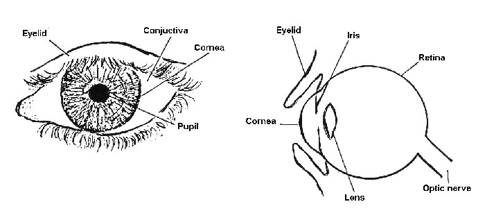
• Know the findings in a normal eye Having taken the HISTORY and measured the VISUAL ACUITY, you can now EXAMINE the eyes. It is important to know the appearances of the NORMAL EYE.
In the NORMAL EYE:
- The eyelids open and close properly.
- The white of the eye (sclera) is white.
- The cornea (the window of the eye) is clear.
- The pupil is black, and becomes small in bright light.
See an illustration of a normal eye in figure 16.5
Figure 16.5: Normal Eye
Examine your patient for abnormalities of the eye:
When examining the eye start with the eyelid, then examine the conjunctiva, the cornea, and finally the pupil in that order. Some of the common abnormalities you should look for in the eyelids, conjunctiva, cornea and pupil are listed below:
Four abnormalities of the EYELIDS:
- Cannot close
- Cannot open
- Eyelashes turn in
- Eyelid is swollen
Four abnormalities of the CONJUNCTIVA:
- It is red
- It is growing onto the cornea
- There is a haemorrhage
- There are white foamy spots (Bitot spots)
Four abnormalities of the CORNEA:
- There is a white scar
- There is a grey spot in a red eye
- There is a foreign body
- There is a laceration
Four abnormalities of the PUPIL:
- It is white
- It is irregular in shape
- It is large and does not become small in bright light
- There is blood in front of it.
If you find that the child has any of the abnormalities mentioned above, refer to hospital.
16.2.2. OPHTHALMIA NEONATORUM:
Ophthalmia neonatorum is a condition in which a baby, in the first twenty-eight days of life, has sticky eye discharge.
Causes
Ophthalmia neonatorum is caused by Neisseria gonorrhoeae (gonococcus) or Chlamydia trachomatis. Have you ever seen and treated a baby with ophthalmia neonatorum? Before you continue reading, stop for a while and do the following activity.
| 10
List three (3) signs and symptoms of a baby with opthalmia neonaturm _____________________________________________________________________ _____________________________________________________________________ _____________________________________________________________________ _____________________________________________________________________
|
Signs and symptoms
A baby with ophthalmia neonatorum presents with the following signs and symptoms:
- Swollen eyelids
- Red and swollen conjunctiva
- Pus discharge from the eye
- Cornea is usually clear, but may show a corneal ulcer
Management
When you see a baby with ophthalmia neonatorum, this is what you should do:
- Clean the eyes with a clean swab and water;
- Apply Tetracycline eye ointment hourly for 4 days, then three times a day for 10 days;
- If the ophthalmia neonatorum is very severe, particularly if there is a corneal ulcer, give also antibiotic eye drops (such as Penicillin or Chloramphenical); every minute for one hour, then every hour for one day, then every 3 hours until there is improvement;
- Give systemic antibiotics (such as Procaine Penicillin 60 mg) by intramuscular injection, or other suitable systemic antibiotic.
Before continuing, do the following activity:
| 11
_____________________________________________________________________ _____________________________________________________________________ _____________________________________________________________________
|
Now confirm your answers as you read the following discussion.
Prevention:
To prevent ophthalmia neonatorum, you should:
- Use a clean swab and water to clean the eyes of each newborn baby immediately at birth, even before the baby has opened his eyes;
- Apply Tetracycline eye ointment once to a new born baby's eyes. If Tetracycline eye ointment is not available, apply one drop of 1% silver nitrate solution.
Next let us look at the third condition of the eye.
16.2.3 XEROPHTHALMIA
Xerophthalmia is dryness of the cornea or the conjunctiva due to Vitamin A deficiency. Xerophthalmia may lead to corneal ulceration and blindness, particularly during the presence of measles infection.
Causes
There are three major causes of Xerophthalmia, these are:
- Malnutrition: Xerophthalmia is caused by a deficiency of vitamin A which may result from:
- Insufficiency of vitamin A in diet (quality of food)
- Inadequacy of amount of food. (quantity of food)
- Inability of the body to use vitamin A
- Depletion of vitamin A from liver stores
Vitamin A can be obtained from two sources. Certain foods, such as eggs, milk, liver and fish provide the vitamin directly to the body. Other foods, such as green leafy vegetables, carrots, tomatoes, mangoes and paw paws contain a substance that can be changed into Vitamin A in the body and then absorbed. A s long as the baby is fed with mother's milk, there is little danger of xerophthalmia. Often, however, when the baby is weaned too early on a diet of rice or gruel he or she may develop Vitamin A deficiency.
Xerophthalmia is likely to occur in malnourished children and often accompanies Protein-Energy-Malnutrition (PEM).
- Malabsorption: Chronic diarrhoea that causes malabsorption of Vitamin A.
- Measles: There is an important relationship between childhood infections and Vitamin A deficiency. When a child becomes sick, he may lose his appetite and not get enough of the vitamin in the food eaten. Such illnesses often reduce the body's ability to absorb and use the vitamin in the food. Without a dietary intake, the vitamin A stored in the liver becomes drained. The diseases most associated with Vitamin A deficiency are measles, whooping cough, round worms and tuberculosis.
Signs and symptoms
A child who has xerophthalmia may present with the following signs and symptoms:
1. Night blindness: The child will be unable to see in dim light, for example in the evening. Mothers are usually quick to recognise the problem as the child no longer moves about in the house or village after dark, but sits in a corner, unable to find his food or toys. Mother's word could be relied upon as an accurate diagnosis when carefully questioned.
2. Bitot’s spots: These spots look like white, sticky foam on the white portion of the eye. They almost always occur in both eyes.
3. Xerosis: Xerosis is dryness of the conjunctiva or cornea.
4. Corneal ulcer: Ulceration of the cornea may develop if the disease is not treated. If you happen to get a child with corneal ulcer in your health unit, this is what you should do:
- Give Vitamin A 200,000 International Units (IU) by mouth on the first day (therapeutic doses).
- On the second day you give Vitamin A 200,000 IU orally again.
- After 3 to 4 weeks you give Vitamin A 200,000 IU orally.
- Apply Tetracycline eye ointment three times a day.
- Apply Atropine eye ointment once a day.
- Apply an eye pad to stop the child rubbing his eyes and causing perforation.
Table 16.2: Vitamin A Supplementation: Source Kenya MOH IMCI Guidelines
| AGE | VITAMIN A CAPSULES | ||
|---|---|---|---|
| 200 000 IU | 100 000 IU | 50 000 IU | |
| Up to 6 months | - | 1/2 capsule | 1 capsule |
| 6 months up to 12 months | 1/2 capsule | 1 capsule | 2 capsules |
| 12 months up to 5 years | 1 capsule | 2 capsules | 4 capsules |
| 12
_____________________________________________________________________ _____________________________________________________________________ _____________________________________________________________________ _____________________________________________________________________
|
Now confirm your answers as you read the following discussion.
Prevention
You can prevent xerophthalmia by:
- Offering nutrition education to parents to prevent malnutrition. Encourage mothers to breastfeed their babies up to 2 years;
- Encouraging mothers to give their children mangoes, pawpaws and green leafy vegetables;
- Giving oral rehydration solution for diarrhoea controls malabsorption;
- Immunizing children against measles;
- Giving Vitamin A capsules to children with measles, malnutrition, malabsorption, or any sign of Vitamin A deficiency, for example, night blindness or Bitot spots. See Table 16.2.2 for dosage.
16.2.4 TRAUMA:
Trauma means injury. There are four main types of injuries to the eye. These are:
- Blunt injuries
- Perforating injuries
- Foreign bodies
- Burns or chemicals in the eye.
The type of injury can usually be ascertained from the history. Remember, you should measure the visual acuity in each eye in all patients with eye problems.
In this section we shall discuss blunt injuries and perforating injuries. Foreign bodies and burns or chemicals in the eye will be discussed in Unit 19.
Blunt Injuries To The Eye
A blunt injury is physical harm or damage due to blunt object. After taking the history and visual acuity, the eye should be examined.
Signs and symptoms: On examination, the patient may have the following signs and symptoms:
- Bruising in the eyelids;
- Fracture of one of the orbital bones.
The clinical signs of an orbital fracture are:
- Bleeding from the nose;
- Anaesthesia of the lower eyelid;
- Double vision;
- Sub-conjunctival haemorrhage due to bleeding under the conjunctiva;
- Abrasion of the cornea, which will present as an acutely painful eye;
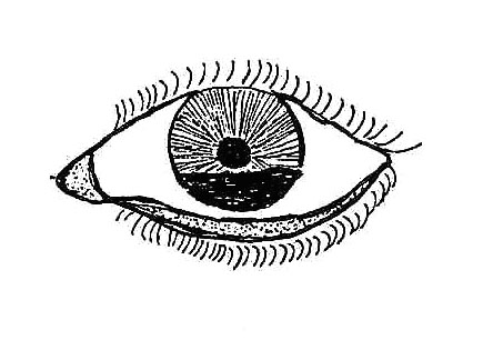
- Distorted pupil or pupil not visible due to haemorrhage.
Fig. 16.6 Haemorrhage in the anterior chamber of the eye due to blunt injury
Management This is how you should manage a child with a blunt eye injury:
- Instruct the mother that the child should have complete bed rest for 5 days.
- Apply antibiotic eye ointment for 5 days;
- Apply eye pad for 5 days;
- Refer to hospital if no improvement after 5 days;
Perforating Injury
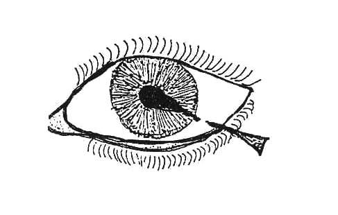
A perforating injury is physical harm or damage due to a sharp object. You often gather this information from taking history and measuring visual acuity. Once you establish that the injury is due to a sharp object that may have resulted in perforation of the eye, it is important to examine the eye very urgently.
Fig. 16.7 Perforating injury to the eye.
Signs and symptoms:
When you examine the eye, the patient may have the following signs and symptoms:
- Lacerations of the eyelids.
- Lacerations of the conjunctiva.
- Injury to the cornea.
- Distorted pupil.
Management: The first aid management of a perforating injury includes:
- Administration of Tetanus Toxoid vaccine.
- Apply antibiotic eye drops to the eye.
- Apply atropine eye drops to the eye.
- Apply eye pad.
Then refer the patient to a hospital immediately.
16.2.5. CONJUNCTIVITIS
Conjunctivitis simply means inflammation of the conjunctiva.
Causes: Conjunctivitis is caused by:
- Bacteria
- Viruses
- Allergy
- Chemicals
- Cosmetics
- Smoke
- Harmful eye practices
Signs and symptoms: A child with conjunctivitis may present with the following signs and symptoms:
- A little pain in the eyes
- Pus or mucus discharge
- Swollen eye lids
- Red conjunctiva
Management: Treatment depends on the cause as follows:
- Bacterial conjunctivitis
- Apply Tetracycline eye ointment three times a day for one week.
- Viral conjunctivitis
- Apply Tetracycline eye ointment three times a day for one week to stop secondary infection.
- Allergic conjunctivitis
- Find out the cause of allergy and remove it.
- Advise the parents to avoid bringing into their house the things to which the child is allergic.
- Advise parents to avoid taking the child where there is something to which the child is allergic.
16.2.6 TRACHOMA
Trachoma is inflammation of the conjuctiva and the cornea caused by Chlamydia trachomatis. It is the most important cause of blindness in the world.
Cause: There are various environmental factors that favour trachoma and various factors that are important in the repeated transmission of trachoma. These are:
- Lack of water;
- Discharge on children's eyes;
- Fingers: Repeated infection with eye - finger – eye;
- Flies: Flies can carry infection from one person's eyes to another's eyes;
- Fomites: Infection can be carried from eye to cloth and from cloth to eye.
| 13
A mother comes to your health facility with a four year old son for treatment. You take the history from the mother, examine the child, and diagnose the child as having trachoma. Why did you conclude that the child has trachoma? What symptoms did he have? _____________________________________________________________________ _____________________________________________________________________ _____________________________________________________________________ _____________________________________________________________________
|
Signs and symptoms: A child with trachoma may present with the following signs and symptoms:
- Red, sore eyes with follicles
- Red conjunctiva with follicles
- Discharge of pus from the eyes
- Conjunctival scars
- Corneal scar
- Blindness after many years
Management:
Trachoma can be managed in the following way:
- Instruct members of the family to wash their faces daily.
- Clean the eyes with cotton swabs and water.
- Apply Tetracycline eye ointment two to three times a day for six weeks.
- In case of severe trachoma, give systemic treatment with tablets of
- Cotrimoxazole or erythromycin 14 days.
- If the visual acuity is poor and if the corneas are not clear then refer to hospital.
Well, we hope you have enjoyed learning about common eye conditions in children. Next we shall discuss common skin conditions.
16.3: COMMON SKIN CONDITIONS
There quite a number of common skin conditions in children, most of which are caused by infection with bacteria, fungi, viruses or invasion of the skin by insects. We hope that by the end of this section you will be able to identify the skin conditions through their signs and symptoms, manage and prevent them effectively.
Before you read on, do the following activity:
| 14
List the common skin conditions that you know: _____________________________________________________________________ _____________________________________________________________________ _____________________________________________________________________ _____________________________________________________________________
|
I believe your answers included some of the following common skin conditions that we see in children:
- Scabies
- Ringworm
- Tropical ulcers
- Atopic Eczema
- Diaper (nappy) rash
- Viral skin infections
- Impetigo
- Pediculosis (louse infestation)
- Jiggers
- Drug eruptions
- Warts and molluscum contagiosum
- Skin tumours such as Kaposi’s sarcoma and Haemangioma.
We shall now discuss each of the skin conditions listed except measles rash, which was discussed in Unit 9. You can refer to thats unit to refresh your mind about it.
16.3.1 SCABIES:
Scabies is a parasitic infestation of the skin with sarcoptes scabiei , a small mite that is not visible to the naked eye. It is characterized by intense itching, papules, vesicles, excoriations and burrows.
Cause
Sarcoptes scabiei, the itch mite, is an arthropod spread by contact with an infested person. The mites burrow into the superficial layers of the skin where they lay eggs. It is this burrowing as well as secretions from the mites that cause severe itiching, particularly during the night.
| 15
List the signs and symptoms of scabies _____________________________________________________________________ _____________________________________________________________________ _____________________________________________________________________ _____________________________________________________________________
|
Now read through the text below and see if your ideas are included.
Signs and symptoms of scabies
Most children with scabies are not brought to seek medical attention. The skin lesions may be so common that parents do not consider them to be a disease.
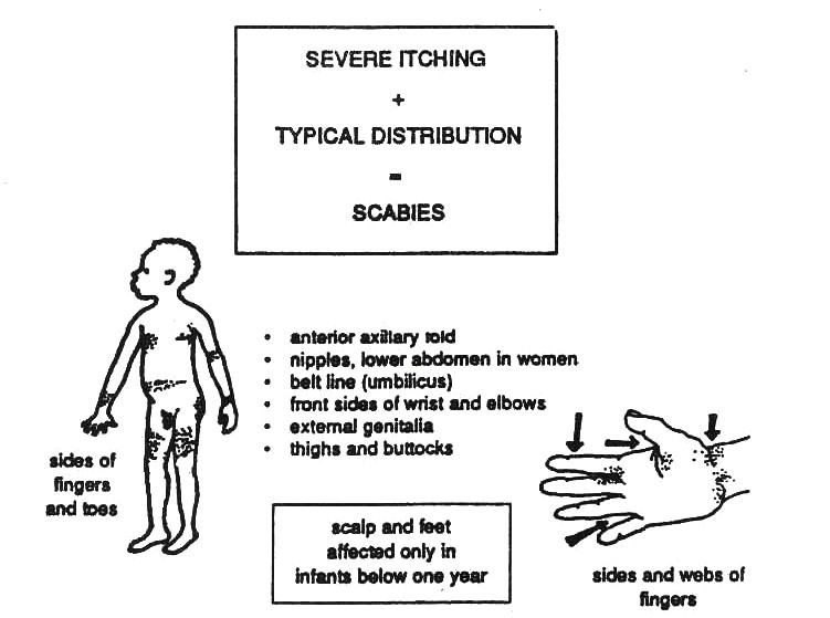
A child with scabies will present to you with skin lesions that intensely itchy, papules, vesicles, excoriations and scaling, the itch being especially at night. The diagnosis is made by finding an itchy rash, very often infected, between fingers an toes, on the writs, buttocks, genitals, arms and legs (see Figure 16.5). In infants, even the head may be infected. Because of scratching, there is usually also bacterial secondary infection.
Treatment
The drug of choice in the treatment of scabies is 25% Benzyl Benzoate Emulsion (BBE). You should give the following instructions to the caretaker of a child with scabies about administration of Benzyl Benzoate Emulsion:
- Give the baby a warm bath.
- Rub a handful of Benzyl Benzoate Emulsion over the whole body, preferably using bare hands.
- Bathe the child after 24 hours and put on clean clothes.
- Protect the baby's eyes while applying Benzyl Benzoate Emulsion on the scalp.
- Repeat the same procedure of application after 4 - 7 days. This is necessary so that you kill the larvae which have hatched since the first treatment.
- Follow the same instruction to treat all the family members.
Fig. 16.8 Common Places for scabies infestation
Complications
A child who has a severe and extensive infestation with scabies and is not treated may develop complications. These complications are the result of secondary bacterial infection. If pustules form and the child develops a fever, you should give antibiotics for five days. Apply Benzyl Benzoate Emulsion when the secondary infection comes down.
| 16
How can you prevent scabies in your community? _____________________________________________________________________ _____________________________________________________________________ _____________________________________________________________________ _____________________________________________________________________
|
Now confirm your answers as you read the following discussion.
Prevention
You can prevent and control the spread of scabies by encouraging members of your community to do the following:
- Wash regularly with soap and water
- Washclothes with soap and water
During your health education talks stress the importance of using soap and water.
16.3.2 RINGWORM (Tinea):
Ringworm is caused by several species of fungi which attack the skin, hair and the nails.
Signs and symptoms
The signs and symptoms of ring worms will depend on which part of the body the infection has occurred:
- Ringworm of the scalp (Tinea capitis): This is the most common one in children. It begins as a small papule that spreads outwards to involve a larger area. Hair in the affected part and it becomes brittle and breaks off easily, leaving characteristic circular bald patches which are itchy.
- Ringworm of the body (Tinea corporis): This is also common in children. It is characterised by flat circular and outward spreading lesions on the body. The ring lesions are reddish, vesicular or pustular and may be dry and scaly or moist and crusted. The central area often clears leaving what looks like normal skin.
- Ringworm of the foot or athlete’s foot (Tinea pedis): A child with ringworm of the foot will present with scaling and cracking of the skin between the toes.
- Ringworm of the nails (Tinea unguium): A child with ringworm of the nails will present with thickening, discoloration and brittleness of one or more of the nails.
Treatment
You should follow the instructions below in the treatment of a child with ringworm infection. Ask the parent to:
- Wash the child's body and clothing with soap and water daily;
- Cut the hair around any ringworm infection on the scalp;
- Apply clotrimazole ointment 8 hourly daily to the affected areas.
Some lesions need treatment for at least 2 weeks, but spontaneous resolution also occurs. You should refer to hospital any children with widespread lesions, lesions resistant to therapy or children with ringworm of the nails.
Prevention
Ringworm and other fungal infections are contagious. You need to prevent them from spreading from one child to other children. Here are some of the ways you can achieve this:
- Do not let a child with a fungal infection sleep with others.
- Do not let different children use the same comb or use each other’s clothing unless these are washed or well cleaned first
- Treat an infected child at once.
16.3.3. TROPICAL ULCERS
There are a few differences between tropical ulcers in children and adults. In children, the ulcers tend to develop more rapidly and become more extensive, but they also heal more quickly.
Cause Tropical ulcers often follow a cut or abrasion of the skin that becomes infected with bacteria, especially in unhygienic settings.
Signs and symptoms A child with a tropical ulcer(s) will present with a rapidly growing necrotic painful ulcer. This ulcer will have raised or undermined edges. The feet and lower legs are the common sites.
Treatment In treating a child with a tropical ulcer, you should do the following:
- Rest the affected leg.
- Clean the ulcer with chlorhexidine solution.
- Excise the necrotic edges.
- Perform daily dressing.
If the above treatment fails, give procaine penicillin 30mg/kg/day for 5 days.
16.3.4 ATOPIC ECZEMA
Eczema is a distinctive inflammatory reaction of the skin
Cause
The cause of eczema is unknown. Constitutional factors or some allergens may sometimes be responsible.
Signs and symptoms
An infant with eczema will present with areas of itchy reddened rash with vesicles, papules, weeping of clear fluid and crusting later, commonly on the cheeks or ears. In older children, the lesions involve the flexural areas of the knees, armpits, elbows and skin folds. A child with chronic eczema will have dry lesions, which may show scaling. Secondary infection occurs.
Treatment
In treating atopic eczema, apply 1% Hydrocortisone skin ointment to the affected area twice a day. In refractory conditions, apply 0.1% Betamethasone ointment twice a day.
If there is secondary infection, add 0.5% chlorhexidine solution or give Phenoxymethyl penicillin tablets 165mg every 6 hours for 5 days. Advise parent to avoid use of soap on the lesion but to wash only with clean water. Other precipitants should also be avoided.
16.3.5 DIAPER RASH
Diaper rash is a rash that appears in the genital area of babies.
Cause
Diaper rash is caused by urine in the baby's diaper (nappy) or bedding that comes in prolonged contact with the skin.
Signs and symptoms
Diaper rash is a very common skin problem in babies. An affected baby will present to you with patches of irritation between the legs or buttocks.
Treatment
You should advise the parent of a baby with a diaper rash to:
- Bath the child daily with lukewarm water and mild soap.
- Keep the baby naked, without diapers and to take the baby out into the sun.
- Change diapers often if they are to be used.
- Use talcum powder only after the rash has gone.
Prevention
In the prevention of diaper rash, you should advise the parents to keep the baby naked (without diapers) and to place the child in the sun for some minutes each day.
16.3.6 VIRAL SKIN INFECTIONS
Viruses cause a wide variety of skin infections in children of all ages. Most viral skin infections clear up spontaneously while others call for treatment that may be specific or non-specific. In Unit 9 of this course, you learned about measles that is caused by a virus and presents with a maculopapular skin rash.
In this section we shall discuss other common viral skin infections that you frequently encounter in your health unit. They are:
- Herpes simplex
- Chicken pox and Herpes zoster
- Molluscum contagiosum
| 17
Describe the presentation of a child with Herpes simplex: _________________________________________________ _________________________________________________ Herpes zoster: _________________________________________________ _________________________________________________ Molluscum contagiosum: __________________________________________ __________________________________________
|
*Herpes Simplex
Herpes simplex, also referred to as Herpes labialis or Herpes febrilis, is a common viral infection of the skin in children. It is usually associated with fever and infections like malaria, pneumonia, meningitis or immunosuppressive diseases like HIV infection.
Signs and symptoms
A child with Herpes Simplex will present with burning crops of clear tense blisters most commonly around the lips, nose, eyes, or external genitalia. The child may have low grade fever.
Treatment
The treatment is generally symptomatic. You should ask the parent of the child to clean the lesion, whether oral or perineal, with antiseptic solution like Chlororhexidene solution 0.5%. You should also treat the underlying disease appropriately.
*Chicken Pox (Varicella) And Herpes Zoster
This is a highly infectious disease caused by virus – varicella zoster virus. Chicken pox and Herpes zoster (shingles) are two different manifestations of the same virus. The virus, whose incubation period is 10-21 days, is spread by aerosol, close contact, infected hands and clothing. In children it is usually a mild disease.
Signs and Symptoms
Various lesions (macules, vesicles, papules and crusts) appear in crops, and are superficial, appearing first on the face, trunk, thighs and upper limbs. The lesions heal completely leaving a healthy, unmarked skin.
Complications
In the newborn or those with depressed immunity, as in HIV infection a vital septicaemia may occur with severe illness. Sometimes a secondary bacterial infection of the spots (rash) occurs. Other complications include varicella pneumonia, hepatitis, encephalitis, etc.
Treatment
- Clean lesions with plenty of water and soap.
- Apply Calamine lotion.
- Give oral antihistamines such as phenergan.
- Penicillin systematically is needed in case of secondary bacterial infection and pneumonia.
*Herpes Zoster (Locally Called Kasipi)
This is caused by the varicella-zoster virus localized in the nerve roots. It may follow an attack of chicken pox.
Signs and Symptoms There is severe pain, followed by erythematous papules which then form vescicles which desicate to form crusts distributed over the area of one or more nerves. Acyclovir 5-8mg per kilogram 8 hourly intravenously indicated in those with generalized infections.
Treatment
- Clean the lesions.
- Relieve pain with pain killers (analgesics).
Note:
Herpes zoster is becoming common, and it is most common among HIV/AIDS infected people.
*Warts and Molluscom Contagiosum
These are fairly common skin infections caused by viruses. Apart from the sometimes unpleasant cosmetic effect, they do not do any harm to the patient, and they usually disappear spontaneously.
Signs and Symptoms
In warts, the horny layer of the skin shows great over-growth to form rough papules with irregular surfaces. Warts commonly occur on the fingers, face, knees, elbows, soles, hands, being either single or multiple.
In molluscum contagiosum, there are small rounded smooth papules that vary in size from that of pin-heads to a pea. They are large and numerous in patients with AIDS. The centre of each lesion is depressed. They contain whitish material. Common sites for this condition include the face, forearms, thighs and genitals.
Treatment
Counselling and reassurance is usually enough. However, if you must treat, then the following are some ways of treatment:
- One drop of phenol on the end of a sharpened matchstick can be applied on isolated lesions.
- Repeated covering with elastoplast strapping can be tried.
- The lesions can be frozen with ethyl chloride and scraped away with a curette.
*Impetigo
Impetigo is a contagious superficial infection of the skin caused by bacteria, usually the bacteria called streptococci or staphylococci.
It is important to know that impetigo is highly contagious and spreads readily between children. Flies are known to transmit the disease.
Note also that complications streptococcal skin infections may cause: Rheumatic fever, acute glomerulonephritis (AGN), septic arthritis, pneumonia, and cellulitis. Therefore, early diagnosis and subsequent treatment is important.
There is a nonbullous and a bullous impetigo. The nonbullous impetigo is an acute superficial skin infection which starts with a small vesicle or pustule that rapidly exudes clear fluid (serum) to form yellow crusted plaques. The lesions most commonly occur on the head, especially the face, but may involve any part of the body. Nonbullous impetigo is common among malnourished children. Its association with tonsillitis or sores in the nose suggests streptococci as the most likely cause.
Caused by Staphylococcus aureus, bullous impetigo is characterized by large loose blisters containing yellow fluid and involving large areas of the body, especially skin folds. It occurs in neonates and young infants. It may be complicated by a fatal septicaemia.
Treatment:
- Remove scabs and crusts with soap and water or half-strength (1.5%) Hydrogen peroxide. Hibitane is good, if available.
- Cut hair away from scalp lesions.
- Apply gentian violet (GV) 1% aqueous solution.
- Penicillin, systemically, is indicated in the following:
- Widespread lesions.
- In all newborns.
- If follow-up is difficult.
- Louse Infestations (Pediculosis)
Lice are transmitted by close, personal contact where hygienic standards are low.
Signs and Symptoms:
- Children are commonly infested with head lice. These parasites cause severe itching. The child scratches the head and this usually causes secondary infection of the scalp. This infection usually spreads to the neck and scapular region.
- Tender lymphadenopathy at the sides and back of the neck may manifest.
- The lice eggs that usually appear as small grey-white specks are attached firmly to the hair.
- Body lice cause small, red, slightly prominent itchy skin lesions when sucking blood. They live and lay eggs in the clothing.
- It is important to note that the louse is an important vector of louse-borne typhus and louse-borne relapsing fever.
Treatment:
The easiest, cheapest and most effective treatment is to shave the head. When this is not possible, do the following:
- DDT 10%, Bezene hexacholoride 1% or Benzyl Benzoate Emulsion (BBE) 25% is applied to the head as powder or solution. The following day, the head should be washed thoroughly and combed with a special nit comb to remove all the eggs.
- When there is secondary infection of the scalp, cut the hair short and apply antibiotic ointment, and give penicillin systemically.
- Look for lice in other members of the family.
- All clothing should be dusted with an insecticide powder.
*Jiggers
The pregnant female sandflea (Tunga penetrans) burrows into the skin, especially on the toes and feet. It ultimately grows to discharge its eggs if not removed. These may hatch out on or in the skin into adult fleas. This causes severe itching and inflammation.
Complications:
Note that tetanus infection can be a complication of jiggers.
Before you read on, do the following activity:
| 18
What factors favour Tunga penetrans infestation _____________________________________________________________________ _____________________________________________________________________ _____________________________________________________________________ _____________________________________________________________________ How would you treat and prevent Tunga penetrans _____________________________________________________________________ _____________________________________________________________________ _____________________________________________________________________ _____________________________________________________________________
|
Now read through the following and check whether your answers are included in the following discussion.
In order to treat and prevent jiggers, the following should be in place:
- Make a correct diagnosis;
- Clean the infected areas properly;
- Remove the flea with a sterile needle;
- Cover the wound with antiseptic gauze;
- You can also ask the mother to remove the jiggers with a fine, clean instrument as she usually does at home. Advise her to clean the child’s feet thoroughly with soap and water before and for 2 days after the procedure;
- Take this opportunity to immunise the child against tetanus if the child is not immunised;
- When there are multiple lesions, apply turpentine, chloroform or kerosene which kill the jiggers with subsequent expulsion by the provoked inflammation.
*Drug Eruptions
Many widely used drugs can produce allergic reactions of the skin as a side effect in susceptible individuals.
First complete the activity below before reading on:
| 19
_____________________________________________________________________ _____________________________________________________________________ _____________________________________________________________________ _____________________________________________________________________
|
I hope your answers included the following drugs known to cause skin reactions:
- Penicillins
- Streptomycin
- Sulphonamides
- Tetracycline
- Barbiturates
Some agents cause skin irritation after direct contact. This is called contact dermatitis. Some of the well-known agents include:
- Iodine
- Pyrethrum
- Baby oils
- Baby powders
Signs and symptoms
There may be a mild, transitory rash or a more severe reaction like urticaria or even generalised eruptions with fever and anaphylactic shock.
Treatment
The best treatment is to try to find out the offending drug and stop using it. Reassess why the drug was given. If the condition (disease) is still there, then use an alternative drug.
- In severe cases, antihistamines are indicated, such as phenergan 1 mg/kg/day, orally in 2-3 divided doses.
- Do not attempt to use antihistimines topically. They sometimes provoke drug eruptions.
|
It is important to note that any drug is dangerous and can cause skin reactions/allergy to susceptible individuals
|
*Skin Tumours
Haemangioma
This is a benign tumor made up of blood vessels that often occurs in newborns. It typically occurs as a purplish or reddish slightly elevated area of skin and may present as a birthmark.
Signs and Symptoms:
Flat or raised vascular tumours of the skin are often seen at birth. Common sites include the face and neck. No treatment is required as these lesions disappear on their own in a few months or years.
Management:
Do not attempt to operate on them. Just reassure the mother. If in doubt please REFER to hospital for assessment by a doctor.
Kaposi’s Sarcoma
This is a malignant tumour of lymphoreticular or vascular cells presenting with nodular skin lesion and diffuse lymphadenopathy. The tumour is rare in children, but with the advent of HIV/AIDS, it is becoming more frequent.
Signs and Symptoms:
The nodular skin lesions usually occur on the limbs. There may be only just thickening of the skin. Sometimes there is swelling of the affected limb and even ulceration in the skin. Nodules may also be found in the palate and the conjuctiva together with periorbital bruising. The lympadeonopathy is diffuse and the lymphnodes are rubbery in consistence.
When it is associated with HIV/AIDS infection, it tends to be widespread and grows very fast. It is a poor sign in HIV/AIDS affected individuals.
Kaposi’s sarcoma should also be suspected in children with persistent diarrhoeal weight loss, intestinal obstruction or abdominal pain
Management:
Refer suspected cases to hospital where they can confirm the diagnosis by carrying out a biopsy and manage it accordingly. It can be treated by surgery or chemotherapy to which it responds well.
SUMMARY
Congratulations! You have now come to the end of this unit. Go through the objectives of this unit which we discussed at the beginning. Have you achieved all of them? If there is any you are not sure of, review the relevant sections. If you feel confident that you have covered them well, then take a break and then complete the attached Tutor Marked Assignment.
Good Luck!!!
|
DIRECTORATE OF LEARNING SYSTEMS DISTANCE EDUCATION COURSES TUTOR MARKED ASSIGNMENT
Name: _________________________________________ Address: _______________________________________ _______________________________________________
CHILD HEALTH COURSE UNIT 16: OTHER CONDITIONS
1. A child is brought to you running a high temperature and with diarrhoea. The parents are convinced that this is due to “false teeth.” a. Give an outline of how you would manage this child:
_____________________________________________________________________ _____________________________________________________________________ _____________________________________________________________________ _____________________________________________________________________
_____________________________________________________________________ _____________________________________________________________________ _____________________________________________________________________ _____________________________________________________________________ _____________________________________________________________________
LIST A ` 1. Enamel____ 2. Deciduous____ 3. Calculus____ 4. Gingivitis____ 5. False teeth____ 6. Plaque ____ 7. Oral thrush
LIST B a. Sticky white or yellow deposit consisting of bacteria. b. Calcified plaque c. Infection caused by candida d. Infection of the bone surrounding teeth e. A disease affecting the gum f. The practice of removing tooth buds in infants g. The hardest tissue of the body h. The first set of teeth in human beings i. Artificial teeth 3. A 16-year-old girl is brought to your health facility by her older sister with an injury on the right eye. You take the history from the girl herself. You examine the injured eye and you decide that the girl has a blunt injury. What made you decide that the girl had a blunt injury but not a perforating injury? _____________________________________________________________________ _____________________________________________________________________ _____________________________________________________________________ _____________________________________________________________________ _____________________________________________________________________
a. What made you conclude that the child has impaired vision? _____________________________________________________________________ _____________________________________________________________________ _____________________________________________________________________ _____________________________________________________________________ _____________________________________________________________________
_____________________________________________________________________ _____________________________________________________________________ _____________________________________________________________________ _____________________________________________________________________ _____________________________________________________________________
5. Robert is a 3 year old boy, with several encrusted blisters on his face, mostly around the mouth. His mother says that the problem all started with a little blister a few days ago and then pus began to come out of it and then more blisters appeared. When you examine the child, you notice that he looks a bit malnourished. There are yellow crusts on his face. a) What do you think is the diagnosis? _____________________________________________________________________ _____________________________________________________________________ _____________________________________________________________________
_____________________________________________________________________ _____________________________________________________________________
Once you complete this assignment, post or bring it in person to AMREF Training Centre. We will mark it and return it to you with comments. Our address is as follows: Directorate of Learning Systems AMREF Headquarters P O Box 27691-00506 Nairobi, Kenya Email: amreftraining@amrefhq.org
|
