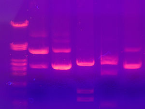
Lab: DNA gel electrophoresis
| DNA electrophoresis is an analytical technique used to separate DNA fragments by size. DNA molecules which are to be analyzed are set upon a viscous medium, the gel, where an electric field induces the DNA to migrate toward the anode, due to the net negative charge of the sugar-phosphate backbone of the DNA chain. The separation of these fragments is accomplished by exploiting the mobilities with which different sized molecules are able to pass through the gel. Longer molecules migrate more slowly because they experience more resistance within the gel. Because the size of the molecule affects its mobility, smaller fragments end up nearer to the anode than longer ones in a given period. After some time, the voltage is removed and the fragmentation gradient is analyzed. For larger separations between similar sized fragments, either the voltage or run time can be increased. Extended runs across a low voltage gel yield the most accurate resolution.
This extract is licensed under the Creative Commons Attribution-ShareAlike license. It uses material from the article "DNA electrophoresis", retrieved 18 Mar 2011. |
Contents
Prep
Learn about how to perform DNA gel electrophoresis by reviewing the concepts presented at the following sites:
- Study the 2nd part of Lab 6: Molecular Biology, DNA Electrophoresis, or the Gel Electrophoresis simulation at The University of Utah's Learn.Genetics
- Read through the full description and explanation of this procedure, which was used as a basis for this lab's protocol: The MacGyver Project: Genomic DNA Extraction and Gel Electrophoresis Experiments Using Everyday Materials
Printout the instructions below for how to create the gel electrophoresis using everyday objects.
Materials Needed
DNA extraction
- Tall narrow container (50mL graduated cylinder, test tube, flower vase)
- Water
- Liquid dish soap or Woolite
- Pipette
- 90% Alcohol (or higher)
- 70% Alcohol
- Living organism: split green peas, wheat germ (uncooked/not toasted), chicken liver, banana, kiwi, oatmeal, yeast, seeds...
- Wooden skewer
- Salt
- Blender
- Strainer
Gel, running buffer and staining
- Plain Agar Agar powder
- Sodium cloride (table salt)
- Sodium bicarbonate (baking soda)
- Distilled water
- pH testing kit
- alkaline buffer
- glycerol (glycerin)
- red food coloring
- pipette
- 2.303% methylene blue (in distilled water)
Electrophoresis box
- Buffer chamber (e.g., large rectangular plastic container)
- Stainless steel wire
- Lego, including a base plate about 5cmx5cm (for building the gel casting chamber)
- Waterproof, moldable plastic sheeting (e.g., Glad Press 'N Seal)
- 5 to 7 9V batteries
- Battery connectors
- Two pairs of mini alligator clip test leads
Procedure
Electrophoresis box
- Run the stainless steel wire along the full length of each short side of the buffer chamber, up the side and over the top, leaving a two cm end for connection to battery set up.
- Label the one side of the buffer chamber positive (anode) and the other negative (cathode).
- Draw an arrow on the chamber from the negative to the positive. (This is the direction in which the DNA will migrate.)
- Use lego bricks to build two sides, three bricks high, onto a square lego base, which will serve as the gel casting chamber. (Note that the square lego base must fit inside the buffer chamber.)
- On the two open sides, run masking tape across to create temporary sides.
- Line the chamber with plastic wrap to create a water tight chamber.
- Create a comb, to be used to create the equally spaced wells in the gel, using a long single-wide length of lego, and adding 3 short single-wide bricks, spaced out across the width of the casting chamber.
DNA sample preparation
- Use one of the procedures used in the DNA extraction lab to obtain the DNA snot.
- Prepare a running buffer with 0.05 g (or 1/64 teaspoon) sodium chloride, 2.7 g (or 1/2 teaspoon) sodium bicarbonate, and distilled water, to final volume of 1 L.
- Set the running buffer’s pH to 7.5, using a pH kit to test and adjust the pH.
- Capture a quantity of DNA snot from the extraction solution.
- Dip the DNA snot into 70% alcohol solution (facilitates removal of excess salts).
- Air dry the DNA for approximately 10 minutes to evaporate residual alcohol.
- Place the dried DNA snot into about 0.5ml (or 1/8 teaspoon) of running buffer and let dissolve overnight at room temperature or a slightly warmer location.
Gel preparation
- Prepare the 1% agar agar gel by dissolving 1.5g (or 1/4 teaspoon) of powdered “Agar Agar” to 100 mL of running buffer and heating the mixture on low heat, with constant stirring, until a homogeneous mixture is achieved.
- Pour the agarose gel mixture into the casting gel chamber until there is a 0.5 cm layer.
- Insert the lego comb into the end of the casting gel chamber, making sure it is inserted evenly, with both ends at the same depth and distance from the end of the chamber.
- Allow 20-30 minutes for gel to solidify.
- Once the gel has solidified, remove the comb and tape very carefully.
DNA sample preparation (continued)
- Prepare a loading solution with 0.5 mL (or 1/8 teaspoon or 10 drops) of glycerol/glycerine, 0.1 mL (1/64 teaspoon or 2 drops) “distilled” water, and several drops of red food coloring.
- For every 0.5 mL (or 1/8 teaspoon or 10 drops) of dissolved DNA sample you have, add 0.05 mL (~1/128 teaspoon or ~1 drop) loading solution. Mix carefully.
Gel running
- Place the gel chamber into the larger buffer chamber oriented so the wells created by the comb are at the end of the chamber with the negative electrode.
- Add running buffer to the buffer chamber until it reaches about 3 mm above the top of the agarose gel.
- Load as much of your sample as you can to each well with a small pipette without letting the wells overflow
- Connect the electrodes and check to make sure the connection is good (by using a multimeter or by checking for bubbles coming up from the metal wires immersed in running buffer).
- Allow the gel to run for approximately 1 hour.
Safety Note: Note that when the gel is running, DO NOT stick your finger in the fluid. There may be enough current flowing to inflict a small shock.
Visualizing the DNA
- Disconnect the electrodes and remove the gel in the gel casting chamber.
- Prepare a 0.02% solution of methylene blue with 0.02 mL (1/128 teaspoon or 1 drop) methylene blue and “distilled” water, to final volume of 1 L.
- Place the gel in the 0.02% solution of methylene blue and allow to stain overnight at room temperature.
- Visualize.
Notes
- ↑ Shirazu, Y., Lee, D., and Abd-Elmessih, E. (2005). The Macgyver Project: Genomic DNA Extraction and Gel Electrophoresis Experiments Using Everyday Materials. Science Creative Quarterly.
