Circulation Worksheet Answers
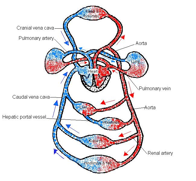
1.The diagram below shows the main vessels of the blood circulation system of a mammal.
- a) Add the following labels to the diagram below:
- caudal vena cava; cranial vena cava; aorta; hepatic portal vessel
- pulmonary artery; pulmonary vein; renal artery
- b) Add arrows to show the direction in which the blood flows
- c) Colour the vessels that carry oxygen rich blood "red" and oxygen poor blood "blue".
2. Arrange the following types of blood vessel in the correct order as blood would flow down them from the heart to the body and back to the heart again.
A. veins; B. venules; C. capillaries; D. arterioles; E. other arteries. F. vena cava; G. aorta
| Heart | Aorta | Other arteries | Arterioles | Capillaries | Venules | Other veins | Vena cava | Heart |
|---|
3. Fill in the blanks in the following table on arteries, veins and capillaries.
| Arteries | Capillaries | Veins | |
|---|---|---|---|
| Structure of wall | 3 layers | 1 cell thick | 3 layers |
| Thickness of wall | Thick | Very thin | Thin |
| Retain shape or collapse
when no blood passes |
Retain shape | Collapse | Collapse |
| Direction of blood flow | Away from heart | From arterioles to venules | Towards heart |
| Speed of blood flow? | Fast | Becoming slower | Slow |
| Blood pressure | High | Decreasing as flows
along capillary |
Low |
| Valves present? | No | No | Yes |
| Pulse present? | Yes | No | No |
| Carry oxygenated/deoxygenated blood? | Oxygenated except pulmonary artery | Blood gives up oxygen as moves along | Deoxygenated except for pulmonary vein |
4.
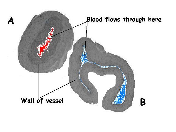
The photo below shows cross sections through an artery and a vein.
- a. Label which vessel is the artery and which the vein. A is the artery and B is the vein.
- b. Give 2 reasons for your answer.
- Reason 1. Assuming these are not the pulmonary artery and vein the vessel with red oxygenated blood is the artery and that with blue deoxygenated blood is the vein.
- Reason 2. The vessel with the thicker wall is the artery.
- Reason 3. The vessel that appears collapsed is the vein as its walls are much thinner than those of the artery.
5.
True or false? If false give the correct answer.
- Mammals have a double open blood system. T
- Arteries only carry oxygenated blood. F The pulmonary artery carries deoxygenated blood.
- Artery walls have many more layers of tissue in them than the walls of veins. F Both arteries and veins have the same 3 layers of tissue in them. The difference is that the layers are much thicker in arteries as they need to withstand the passage of high pressure blood.
- The pulse is only felt in arteries. T
- Arteries have valves in them to stop the blood flowing backwards. F Veins have valves as there is no pulse and the blood is at low pressure so there is a tendency for the blood to pool or flow backwards.
- Blood leaks out of capillaries so that the oxygen and glucose etc. can reach the cells. F Many substances in the blood pass through the capillary wall to enter the tissue fluid that surrounds the cells. These substances move across by various processes but the blood can not be said to leak out of undamaged capillaries.
- Diastole is the phase between pulses. T
- Blood flows along veins back to the heart because of gravity. T Gravity may help blood flow in veins located above the heart but generally most of the movement of blood in veins is brought about by contraction of the surrounding muscles.
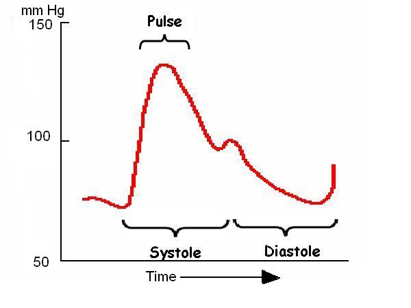
6. The diagram below shows the changes in the blood pressure during the passage of the pulse along an artery.
- Add labels to the diagram to show: the pulse; diastole; systole.
7. As there is no pulse in veins, what moves the blood along them? (Give at least 2 methods)
- 1. The contraction of the muscles surrounding the veins.
- 2. Gravity.
- 3. The contracting heart pulls blood along the veins towards it.
8. Name the vessel that:
- Carries oxygenated blood to the heart muscle. The coronary arteries.
- Supplies the brain with oxygenated blood.The carotid artery.
- Carries deoxygenated blood to the lungs.The pulmonary artery.
- Carries blood from the intestines to the liver. The hepatic portal vessel.
- Carries deoxygenated blood away from the kidneys.The renal vein.
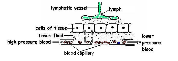
9. Look at the diagram below and then answer the following questions.
- a. Which is the blood capillary? See diagram.
- b. How thick is the wall of the capillary? Very thin -one cell thick.
- c. What is happening to the blood pressure as the blood flows along the capillary? It decreases.
- d. What substances pass out of the capillary walls to surround the tissues? Water, oxygen, salts, amino acids, glucose, etc.
- e. What is tissue fluid? It is the clear fluid that surrounds the cells of the tissues It is formed from blood plasma.
- f. Which vessel is the lymphatic vessel? See labeled diagram.
- g. How do lymphatic vessels differ from capillaries? They are similar in structure having walls only one cell thick. The main difference is that capillaries carry blood whereas lymphatic vessels carry lymph.
- h. What passes into the lymphatic vessel? Tissue fluid passes into lymphatic vessels and (Hey Presto!) it becomes lymph
- i. How does lymph differ from tissue fluid? Both have a similar constitution. The main difference is one of geography. Tissue fluid surrounds the tissues while lymph flows in lymphatic vessels.
- j. Why does the fluid leave the capillary at the beginning of the capillary bed and flow back in at the other end? This is quite a complicated question probably above the level required but here goes. The tissue fluid leaves the capillary at the arterial end because it is forced out by the high blood pressure. At the other end of the capillary the blood pressure is lower and because water has left the capillary at the arterial end the blood in it is more concentrated. This means that the water outside the capillary is “pulled” into it by osmosis. Click here to find out more.


