Bacterial Structure
This is based on New Zealand Qualification Authority Unit 8024 entitled "Demonstrate knowledge of bacterial structure."
To view a power point on this unit follow this link [1]
Contents
1.1 Chemical Composition and Ultrastructure in Cell Wall
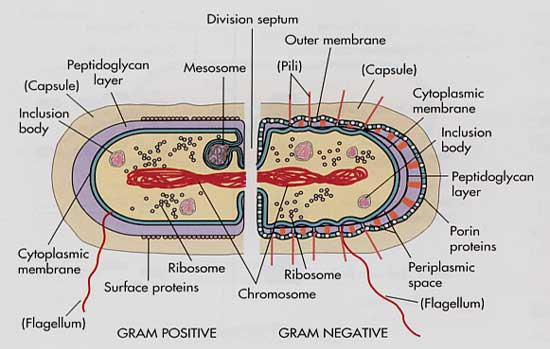
The comparison identifies the similarities and differences in the chemical composition and ultrastructure in the wall of the two types of bacteria.
All bacteria have a cell membrane made up of phospholipid bilayers, which separates the internal contents of the cell from the external environment by creating a semipermeable barrier. Lipids are amphiphilic molecules [Reference: http://en.wikipedia.org/wiki/Amphiphilic], the bilayer is composed of two layers of amphiphilic molecules bound by hydrophobic effect.Usually bacterial cell membranes differ from eucaryotic membranes in lacking sterols such as cholesterol and contain penta-cyclic sterol like molecules are called hopanoids which stabilize the bacterial membrane.
The cell membrane regulates particles entering and exiting the cell, which are used or produced by the cell, during synthesis. Without this type of cell regulation, chemicals toxic to the cell could freely enter, causing cell dysfunction or death.
All cells eukaryotic and prokaryotic have this type of cell regulation; some bacteria though have the increased protection of a peptidoglycan layer,an example being Staphylococcus. Other bateria have a thin peptidoglycan layer, and an outer membrane,an example being Escherichia coli. Bacterial cells that have a thick peptidoglycan layer are classified as Gram positive and the bacteria with a thin peptidoglycan layer are Gram negative.
Some bacterial cells can also gain extra protection from a thick slime capsule.
Comparison of Gram-Positive and Gram-Negative cell walls:
| Characteristic | Gram-Positive | Gram-Negative |
|---|---|---|
| Number of major layers | 1 | 2 |
| Chemical Composition | Peptidoglycan , Techoic acid ,Lipotechoic acid | Lipopolysaccharide ,Lipoprotein ,Peptidoglycan |
| Overall thickness | Thicker(20-80 nm) | Thinner(8-11 nm) |
| Outer Membrane | No | Yes |
| Periplasm Space | Narrow | Extensive |
| Porin Protein | No | Yes |
| Permeability to molecules | More Penetrable | Less Penetrable |
This link will take you to Cells Alive, to see cell animations.[2]
For pictures of Gram positive vs Gram negative. [3] Slides 22 and 23.
| PROCARYOTIC BACTERIA | EUCARYOTIC BACTERIA |
|---|---|
| 1. Heat resistant spores are formed by some species(Endospores). cell 1 | No |
| 2. Poly unsaturated fatty acids or sterols are rare in the membrane. | Common. |
| 3. Some species fix atmospheric nitrogen and able to dissimilate Nitrate to nitrogen gas. | No. |
| 4. Some species uses inorganic compound as sole energy source. | No. |
| 5. Ribosomes are dispersed through out cytoplasm. | To endoplasmic reticulum. |
| 6. Mitochondria and chloroplast is absent. | Present. |
However the differences between cell walls aren't limited to just these two charecteristics of gram staining for instance photosynthetic bacteria may be filled with tightly packed folds of their outer membrane. The effect of these creased membranes is to increase the surface area on which photosynthesis can take place.
Mollicutes have no cell wall at all and are usually parasitic by nature. These cells are very small and usually contain sterols to add rigidity to the cell membrane.
1.2 Gram Staining Comparision
The comparison relates the Gram stain characteristics to the chemical compositions and physical composition of the cell
Bacteria can be separated into two groups determined by the type of peptidoglycan outer layer. These two groups are called gram positive and gram negative.
Gram stains are used to enhance the features of specific cell constituents such as endospores, nuclear regions and granules. In Gram stain dyes like crystal violet, iodine solution and safarnin are used. They are positively charged and combine with negatively charged and fixes dyes inside the cell. The cell components such as nucleic acid and acidic polysaccharides acidic dye have an affinity for positively charged constituents like proteins etc. Thus bacterium that have a deep cell wall made of peptidoglycan (carbohydrate polymers cross-linked by protein) will retain a purple colour when stained with crystal violet solution and are known as Gram-positive. Other bacteria have double cell walls with a thin inner wall of peptidoglycan and an outer wall of carbohydrates, proteins, and lipids. Such bacteria do not stain purple with crystal violet and are known as Gram-negative. [4]
The type of peptidoglycan layer also determines the shape of each type of bacteria.
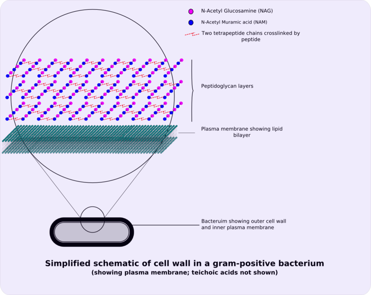
In gram positive bacteria the outer layer of peptidoglycan is relatively thick and has no outer membrane. It is this outer layer that becomes stained purple in the gram staining process, and so is called gram positive.
Gram-positive cellwall
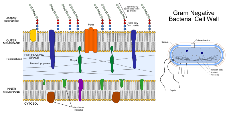
In gram negative bacteria the peptidoglycan layer is relatively thin, but the cell also gains protection through an outer membrane. This membrane covered thin layer of peptidoglycan does not retain the purple stain in the gram staining process, so these bacteria are known as gram negative.
Link to Wikipedia main article on gram staining [5].
http://www.youtube.com/watch?v=OQ6C-gj_UHM
1.3 Major External Structures
The comparison explains the composition and biological properties of major external structures.
Capsule
Bacteria secrete extracellular polymers outside of their cell walls. These polymers are usually composed of polysaccharides and sometimes protein. Capsules are relatively impermeable structures that cannot be stained with dyes such as India ink. They are structures that help protect bacteria from phagocytosis and desiccation India ink (or Indian ink in British English), also called Chinese ink since it may have been first developed in either China or India, is a simple black ink once widely used for writing and printing, and now more commonly used for drawing, especially when inking comics and comic strips. Indian ink generally will readily clog the fountain pens if not used for long time, its then necessary to use water to unclog it. An exception to this is Pelikan Fount India, which does not contain shellac, the substance which can cause clogging.
A capsule is a well organized but easily washed off layer outside bacteria.eg;A Slimelayer which is a zone of diffuse material tht is unorganized and can be easily removed.The capsules and slime layers forms a network of polysaccharides called as glycocalyx extending from the surface of the bacteria to the other cells.But capsules can be construted of non-glucose substances.eg;Bacillus anthracis has a capsule of poly-D-glutamic acid.Mainly archaebacteria contain glycoprotien made capsules called S layers which promotes the cell adhesion to surfaces.
A capsule made up of polysaccharides (complex carbohydrates). Capsules play a number of roles, but the most important are to keep the bacterium from drying out and to protect it from phagocytosis (engulfing) by larger microorganisms. The capsule is a major virulence factor in the major disease-causing bacteria, such as Escherichia coli and Streptococcus pneumoniae. Nonencapsulated mutants of these organisms are avirulent, i.e. they don't cause disease. [6]
Flagella
Bacteria can be motile or non motile, which if they are motile means they can freely move in a desired direction, beneficial to that bacterium. To be motile bacteria can have one, two or many flagellum, depending on the type.
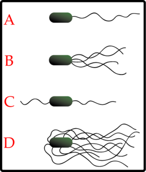
Examples of bacterial flagella:
A - Monotrichous
B - Lophotrichous
C - Amphitrichous
D - Peritrichous
Flagella are rigid structres about 20nm across and 15-20um long.Flegella has three parts.1- the longest portion is the filament which is a hollow rigid cylinder made of the protien called Flagellin.2-the basal body which is embedded in the cell.3- hook a short,curved segment links the filament to its basal body and acts as a flexible coupling.
Flagella are made from a single type of protein and are attached to the outside of the cell. They can be attached at the end, in the middle, or all over the cell as shown in the diagram above. The flagella is helitical and acts as a propeller. It can turn clockwise or anti clockwise to propel the bacteria in different directions. A protein rings in the cell's membrane that act as bearing to aid rotation.
There are motile gram positive and gram negative bacteria.
Pili
Pili sometimes referred as fimbriae are the short, fine, hairlike apendages or projections in both gram negative and positive bacteria.
They are thinner than flagella and not involved in bacterial motility, although some sources disagree with this:
[7].Some pili, designated type IV pili, generate motile forces. The external termini of the pili adhere to solid substrate, either the surface to which the bacteria are attached or to other bacteria, and subsequent pilus contraction pulls the bacteria forward, not unlike a grappling hook. As type IV pilus-mediated movement is typically jerky, it is called twitching motility, as distinct from other forms of bacterial motility, such as are mediated by flagella.
Some types of bacteria uses pili or fimbriae to attach themselves to solid surfaces such as rocks in streams and host tissues. These pili also allow some bacteria to recognize (using lectins to adhere to target cells because they can recognize oligosaccharideOligosaccharide units on the surface of these target cells), stick to, and invade cells linings [8].
Pili are sometimes used to connect the bacterium to another of its species, or to another bacterium of a different species, and build a bridge between the cytoplasms of either cells. All pili are primarily composed of oligomeric pilin proteins that enables the transfer of plasmids between the bacteria. Some bacterial viruses or bacteriophages attach to receptors on sex pili at the start of their reproductive cycle. Despite its name, the sex pilus is not used for sexual reproduction.
Attachment of bacteria to host surfaces is required for colonization during infection or to initiate formation of a biofilm. A fimbria is a short pilus that is used to attach the bacterium to a surface. Fimbriae are found in both Gram-negative and Gram-positive bacteria. In Gram positive bacteria, the pilin subunits are covalently linked.
Lipopolysaccharides
Lipopolysaccharides (LPS) are large molecules consisting of a lipid and a polysaccharide joined by covalent bond. They are found in gram negative bacteria. They are contributing to the structural integrity of bacteria and protecting the membrane from certain kinds of chemical attacks. It also increase the negative charge of the cell membrane and help to stabilize the whole membrane structure.
Composition, the cell structure comprises of three parts:
- Polysaccharide side chains
- Core polysaccharide
- Lipid A
Other than contributing to the negative charge on the bacterial surface, LPS can act as an Endotoxin because of the toxic characterof the lipid A.Eventhough it allows the passage of glucose and monosaccharides through the special permeable Porin proteins but blocks passage of antibiotics and bile salts.
Mycolic Acid
Mycolic acids are found in the cell walls of mycolata taxon [9]. The acid fastness of mycobacterium is attributed to certain high molecular weight lipids. They are lipids connected to long chain to fatty acids.
Mycolic acids can be used as an identification aid as each is composed of 60 to 90 carbon atoms and the exact number varies by species.
2.1 Composition and Function of Bacterial Structures
The description outlines the composition and function of bacterial structures.
Cell Structure
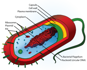
Generic prokaryote cell
CAPSULE
The capsule are made of extracellular polymers composed of polysaccharides and in some cases proteins.
They are made mainly to protect itself from dehydration and being engulfed by bigger micro-organisms although has other purposes as well and is a fairly impermeable structure with dyes like India ink not holding colour.
The encapsulation is the main reason the major disease-causing bacteria are so effective. Through their surface polysaccharide capsules bacteria cloak protein structures on their surfaces to which antibodies could be directed to protect the host. Generally, humans can generate an effective antibody responses against bacterial polysaccharide capsules, but this ability is diminished at extremes of age, so that infants and the elderly are particular prone to invasive infection with encapsulated pathogens. [10]
RIBOSOME
Ribosomes consist of a small and large subunits made up of a complex proteins and RNAs . These are the sites of protein synthesis in the cell. The Ribosome’s in prokaryotic cells although similar in shape and function to those of eukaryotic cells are in different in the nature of the proteins and RNAs that make up their structure. This have provided to be very useful to the human population as antibiotics that acts by inhibiting protein synthesis in bacteria are but not effective against eukaryotic protein synthesis, thus allowing selective toxicity. Archaebacterial ribosome’s [Reference: http://en.wikipedia.org/wiki/Archaea] are the same as those of the eubacteria but, in some features they are similar to eukaryotic ribosome’s in that they are resistant to antibiotics like streptomycin and chloramphenicol, and sensitive to the action of diphtheria toxin.
Ribosomes present in the matrix synthesize protiens to remain with in the cell,whereas the ribosomes present in the plasma membrane make protiens for transport ot outside.Procaryotic ribosomes are smaller than eucaryotic ribosomes and are called as 70S ribosomes.They have dimensions of 14-15nm by 20nm and having a molecular weight of 2.7million. 70S ribosomes constitute of two subunits 30S and 50S repectively,but the total sum of the two subunits give 80S ribosome which is present in eucaryotic cells.The S stands for Svedberg units ,which is the sedimentaion coefficient.
THE NUCLEOID
The nucleoid is an area of the cell where genetic material is localised.
The procaryotic chromosome is a single circle of double stranded DNA, located at in an irregularly shaped region called the nucleoid. It is otherwise called as nuclear body or chromatin body. Nucleoid is composed of 60% DNA, some RNA and a small amount of protien. The DNA is coiled and looped extensively with the aid of nucleoid protiens but not like the histone protiens chromatins in eukaryotic cells.
Diagram of differences between eukaryotic and procaryotic nucleoids: [11]
PLASMIDS
Plasmids are extra chromosomal DNA capable of independant replication and can transfer between cells during recombination.
Plasmids are approximatly tenth the size of prokaryotic chromosomes and are stable. These stable extra chromosomal DNA molecules exist as closed loops containing 5 to 100 genes. There can be one or more plasmids in a cell that may contain similar or different genes. Not all cells contain plasmids, they are not essential for cellular growth but provide a level of genetic flexibility.
Many bacteria possess plasmids which are the circular double stranded DNA molecules in addition to their chromosomes.These plasmids can exist and replicate independently of the chromosome or may be integrate with it and they are inherited or passed on to the progeny. Plasmid genes can render bacteriadrug resistant, give them new metabolic abilities, can make them pathogenic, or endow them with some other properties.
These plasmids are widely used in Recombinanat DNA technology. This comes about through their unique parasitic and symbiotic behaviour with other species. In the case of Rhizobia for example it is able to invade the cytoplasm of root cells and then transcribes over twenty different nitrogen fixing genes. Propperly functioning symbiosis allows the plant nitrogen for growth.
MESOSOME
Mesosome is the common membranous structure in bacteria as bacterial cytoplasm lacks complex organelles like mitochondria or chloroplasts.Mesosomes are the invaginations of the plasma membrane in the shape of vesicles, tubules ,or lamellae.They are seen in both gram positive and gram negative bacteria.It is a real fact that the exact function of the mesosome is yet to be found.They arev beleived to be involved in the cellwall formation during division and in chromosome replication;as they are normally found next to the septa or cross cellwalls in the dividing cells.Presently new researches shows that mesosomes are artifacts generatd during the chemical fixation of bacteria for electron microscopy,and they represent the parts of the plasma membrane which is chemically different and more disrupted by the chemical fixatives.
CYTOPLASMIC MEMBRANE
The cytoplasm is the contents of a cell that is enclosed within the plasma membrane. In eukaryotic cells the cytoplasm contains organelles, such as mitochondria, that are filled with liquid kept separate from the cytoplasm by cell membranes. The part of the cytoplasm that is not held within organelles is called the cytosol. The cytosol is a complex mixture of cytoskeleton filaments, dissolved molecules, and water that fills much of the volume of a cell. The cytosol is a gel, with a network of fibers dispersed through water. Due to this network of pores and high concentrations of dissolved macromolecules, such as proteins, an effect called macromolecular crowding occurs and the cytosol does not act as an ideal solution. This crowding effect alters how the components of the cytosol interact with each other.
• 1 Constituents
o 1.1 Cytosol
o 1.2 Organelles
o 1.3 Cytoplasmic inclusions
Cytosol
Proteins in different cellular compartments and structures tagged with green fluorescent protein. The cytosol is the portion of a cell that is not enclosed within membrane-bound organelles. The cytosol is a translucent fluid in which the other cytoplasmic elements are suspended. Cytosol makes up about 70% of the cell volume and is composed of water, salts and organic molecules. The cytoplasm also contains the protein filaments that make up the cytoskeleton, as well as soluble proteins and large structures such as ribosome’s, proteasomes, and the mysterious vault complexes. The inner, granular and more fluid portion of the cytoplasm is referred to as endoplasm.
Organelles
Organelles are membrane-bound compartments within the cell that have specific functions. Some major organelles that are suspended in the cytosol are the mitochondria, the endoplasmic reticulum, the Golgi apparatus, lysosomes, and in plant cells chloroplasts.
Cytoplasmic inclusions
The inclusions are small particles of insoluble substances suspended in the cytosol. A huge range of inclusions exist in different cell types, and range from crystals of calcium oxalate or silicon dioxide in plants, to granules of energy-storage materials such as starchs, glycogen, or polyhydroxybutyrate. A particularly widespread example are lipid droplets, which are spherical droplets composed of lipids and proteins that are used in both prokaryotes and eukaryotes as a way of storing lipids such as fatty acids and sterols. Lipid droplets make up much of the volume of adipocytes, which are specialized lipid-storage cells, but they are also found in a range of other cell types.
Mycolic Acid
Mycolic acids are long chain saturated fatty acids (60 - 90 carbons long) and are found in the cell walls of nocardiae, corynebacteria and mycobacteria, where it makes up to 60% of the cell wall, as well as in waxes and gycolipids.
Due to the mycolic acid being a lipid any cells containing the acid do not stain well using the Gram stain techniques so instead heat and a solvent such as phenol are used. Once stained the cells hold the stain despite being washed with mineral acids and alcohols. This classifies them as acid fast bacteria.
For a diagram of the mycobacterial cell wall please use the following link: [12]
Periplasm
In gram negative prokaryotes the plasma membrane is separated from the outer membrane by a gap called Periplasm. In periplasm, there are 50 kinds of proteins. Most of them are hydrolytic enzymes and binding proteins. The main function of periplasm is to set apart enzymes and prevent them from leaving the cell.
3.1-3.2
The description relates their chemical and structural properties to the resistance of endospores.
Endospores
Endospores are formed as a genetic survival capsule from within bacteria, a dormant cryptobiotic cell called an endospore, at times of environmental stress and are made up of DNA and ribosomes in cytoplasm within a very hard protective "shell". Full diagram: [[13]]
Endospores are highly resistant to environmental stresses and can remain viable indefinitely under appropriate environmental conditions and even in extreme conditions, such as extreme temperatures and in the absence of water which would kill most living things. This survivability is thought to be due to the extremely low water content of endospores. Once the environmental pressure is relieved however they germinate and become vegetitive cells. [[14]]
Other bacteria use different spore forming processes, such as cyst and heterocyst formation, which can also increase the survival rate of these bacteria. These dormant forms have limited resistants to extreme conditions in comparison to endospores.
Predominantly only gram positive bacteria produce endospores, of which Bacillus and Clostridium are the most common.
Endospores are formed by vegetative bacterial cells in a process called sporillation. Only one spore can be formed per bacterial cell, which can be formed in about eight hours and remain dormant for many years until the right environment triggers the cell to become active again.
Some Bacteria make exospores or cysts instead of endospores but are not as resistant to harsh conditions.
To watch step by step endospore formation please use following link:http://www.microbelibrary.org/images/Gauthier/Endopore_formation.htm

Endospores are green, vegetative (non endospore bacteria) cells are red.
FACTORS LEADING TO THE SPORE FORMATION
Endospores are the special resistant, dormant structures formed by many vegetative cells of gram positive bacteria. Endospores are extraordinarily resistant to environmental stresses like heat, ultraviolet radiation, chemical disinfectants and desiccation. This endospore formation makes a bacteria a dangerous pathogen. The position of the spore in the bacterialcell is called as sporangium and it is often covered by a delicate covering called exosporium. About 15% of the spores dryweight is dipicolinic acid complexed with calcium ions which is not contributing to the heat resiatance.
Sporogenesis or spore formation normally starts when bacterial growth ceases due to the lack of nutrients. Sporogenesis is a seven stage process: 1-The nuclear material forms an axial filament. 2-The cell membrane gets folded inwards to enclose the part of the DNA; this completes the immature spore. 3-The membrane grows to engulf the immature spore in asecond membrane. 4-A cortex is formed in the space between the two membranes and both calcium ions and dipicolinic acid gets accumulated. 5-Protien coats are formed around the cortex. 6-The spore matuarion occurs. 7-The lysis of sporangium and the spore liberated. This entire process requires only 10 hours in Bacillus megaterium.
CHEMICAL AND STRUCTURAL PROPERTIES CONTRIBUTING TO THE RESISTANCE OF SPORES
The cortex beneath the spore coat and the spore is made of peptidoglycan and the dipicolinic acid complexed calcium ions stabilizes the spore nucleic acids. Very specialized small, acid soluble DNA-binding protiens present in the endospore saturate the spore DNA and protect it from heat, radiation, desiccation and chemicals. When the cortex move water from the protoplast by osmosis, the protoplast is totally dehydrated, hence protecting it from both extremes of heat and radiation (?).
In fact, the factors contributing towards the heat resistance of the endospores are calcium-dipicolinate and acid soluble protien stabilization of DNA, protoplast dehydartion and bacterial adaptation to high temperatures repectively. But endospores can be killed by autoclaving or burning.
Reactivation of the endospore occurs when conditions are more favourably suited to the bacterial cell involved. The process involves the three steps: activation, germination, and outgrowth. Even in optimal conditions the bacterial cell can fail to germinate unless activation occurs. Activation can be triggered by heating an endospore gently. Germination involves breaking hibernation by triggering its metabolic activity and is commonly characterised by swelling of the endospore, an increase in metabolic activity, rupture or absorption of the spore coat, and loss of resistance to environmental stresses. Outgrowth follows germination and involves the endospore core making new chemical components and shedding the old spore coat to develop into a fully functional vegetative bacterial cell which has the ability to divide and reproduce.
References
Bacteria. (2008). In Encyclopædia Britannica. Retrieved September 22, 2008, from Encyclopædia Britannica Online: [http://www.britannica.com/EBchecked/topic/48203/bacteria ]
'Instant Notes In Microbiology (1999) BIOS Scientific Publishers, J.Nicklin, K. Graeme-Cook, T.Paget & R. Killington'
Retrieved from "http://wikieducator.org/User:Radhey"
Image:Average prokaryote cell- en.svg
From Wikipedia, the free encyclopedia. Retrieved September 22, 2008, from [15]
Image:Gram-positive cellwall-schematic.png
From Wikipedia, the free encyclopedia. Retrieved October 30, 2008, from [16]
Image:Gram negative cell wall.svg
From Wikipedia, the free encyclopedia. Retrieved September 30, 2008, from [17]
Alcomo's Fundamentals of Microbilogy
Microbiology by J.Nicklein, K.graeme-Cook, T.Paget &R. Killington
MICROBIOLOGY by LANSING M PRESCOTT,JOHN P HARLEY,DONALD A KLEIN
